Day 1 :
Keynote Forum
Sanjay Gandhi
Southmead Hospital, UK
Keynote: The role of computer-assisted detection (CAD), artificial intelligence and machine learning in the medical imaging data overload problem: Current practice, limitations and new developments
Time : 09:45-10:35

Biography:
Sanjay Gandhi in his 30-year career spanning over the UK, India and the USA, has won several outstanding awards for his contributions to the Healthcare Innovations and Education. Times TV Network honoured him as a Global Academic Icon and the British Medical Association honoured him as a Highly Commended Editor. He has published 8 textbooks and written several papers on the use of cutting-edge technology to improve patient care. He has expertise in innovative Telehealth products including CAD, Diagnostic & Assessment Tools, PACS, etc. and has been an advisor to multinational organisations. He has also been involved in numerous research projects and collaborative trials. He has regularly published accomplished articles, rare medical conditions and research papers in reputable indexed international journals such as British Journal of Radiology (BJR), British Journal of Hospital Medicine (BJHM) and British Medical Journal (BMJ). He is on the Editorial Boards of four peer-reviewed medical journals. His current research areas are Computer-Assisted Detection (CAD) to find polyps on CT colonography and he is supervising PhD Research on the use of Artificial Intelligence and Machine Learning.
Abstract:
The demand for diagnostic imaging has continued to increase dramatically over the past few decades. More than 5 billion diagnostic tests are performed globally each year. Replacement of older modalities such as Barium enema with CT colonoscopy and isotopeVQ-scans with CT pulmonary angiography has also massively contributed to increased image datasets per examination. As a result, the majority of Radiologists and other healthcare professionals have to review tens of thousands of images every day. Fortunately, technologies such as Computer Aided Detection (CAD), 3D processing and automated image analysis have also continued to develop. These are becoming increasingly more reliable and affordable. Computer-assisted polyps and cancers detection on virtual-colonography, nodules on lung cancer screening and analysis of breast lumps on MR mammography are just a few of the examples. We will discuss the accuracy and use of different CAD programmes. Our research has shown that a large variation exists in sensitivity and PPV of commercially available software. Some programmes suffer from very long analysis times. Hence, companies producing CAD tools need to address these issues. The role and potential of new technologies such as Artificial Intelligence (AI) and machine learning in coping with the massive increase in the medical imaging workload will be explored. This talk will cover the pitfalls and provide practical tips on the use of these techniques. Such information is useful for Radiologists and Radiographers/Technicians. In addition, CAD developers and other healthcare sector’s entrepreneurs might find this discussion useful in order to develop future products.
Keynote Forum
Vikas Leelavati Balasaheb Jadhav
Dr.D.Y.Patil University, India
Keynote: Trans abdominal sonography of the stomach & duodenum
Time : 10:55-11:45

Biography:
Vikas Leelavati Balasaheb Jadhav has completed Post-graduation in Radiology in 1994. He has 23 years of experience in the field of Gastro-intestinal tract ultrasound and diagnostic as well as therapeutic interventional sonography. He has four Indian patents and an international patent in the field of Gastro-intestinal tract sonography and Radiology, since 2008. He has delivered many lectures in Indian as well as international conferences in nearly 27 countries as an Invited Guest Faculty, since 2000. He is a Consultant Radiologist and Specialist in Unconventional Gastro-Intestinal Tract Ultrasound and Diagnostic as well Therapeutic Interventional Sonologist in Pune, India.
Abstract:
Trans-abdominal sonography of the stomach and duodenum can reveal following diseases: gastritis, duodenitis, and acid gastritis. An ulcer, whether it is superficial, deep with risk of impending perforation, perforated, sealed perforation, chronic ulcer and post-healing fibrosis and stricture, polyps and diverticulum, benign intra-mural tumors, intra-mural haematoma, duodenal outlet obstruction due to annular pancreas, gastro-duodenal ascariasis, pancreatic or biliary stents, foreign body, necrotizing gastro-duodenitis, tuberculosis, lesions of ampulla of vater like prolapsed, benign and infiltrating mass lesions. Neoplastic lesion is usually a segment involvement, and shows irregularly thickened, hypoechoic and aperistaltic wall with loss of normal layering pattern. It is usually a solitary stricture and has eccentric irregular luminal narrowing. It shows loss of normal gut signature, enlargement of the involved segment seen, and shouldering effect at the ends of stricture is most common feature. Enlarged lymph-nodes around may be seen. Primary arising from wall itself and secondary are invasion from peri-ampullary malignancy or distant metastasis. All these cases are compared and proved with gold standards like surgery and endoscopy. Some extra efforts taken during all routine or emergent ultrasonography examinations can be an effective non-invasive method to diagnose primarily hitherto unsuspected benign and malignant gastro-intestinal tract lesions, so should be the investigation of choice.
- Radiography | Image Proceesing | Imaging Technology
Location: Sunset -1

Chair
Abdulrahman A. S. Alsayyari
Qassim University, Saudi Arabia
Session Introduction
Abdulrahman A. S. Alsayyari
Qassim University, Saudi Arabia
Title: Study of common requested radiographs and relative exposure dose in Qassim province

Biography:
Abdulrahman Alsayyari, is a vice dean of the college of applied medical sciences at Qassim university. He obtained his PhD degree from university of Queensland at Australia. He worked in both sector clinical and educational where he developed his experience and knowledge to improve the healthcare services for the Saudi population.
Abstract:
The objective of the article was to study the common requested radiographs and relative exposure dose in Qassim province in Kingdom of Saudi Arabia. The method was retrospective and analytical study for collected variables as radiographs, relative entrance surface dose (ESD) and the effective dose, patient age, gender and causative factors. The doses have been derived from the product of system output, mAs, back scatter factor BSF, focal detector distance FDD and focus – skin- distance FSD based on the equation stated by ICRU, (2005) and Davies et al, (1997):
The objective of the article was to study the common requested radiographs and relative exposure dose in Qassim province in Kingdom of Saudi Arabia. The method was retrospective and analytical study for collected variables as radiographs, relative entrance surface dose (ESD) and the effective dose, patient age, gender and causative factors. The doses have been derived from the product of system output, mAs, back scatter factor BSF, focal detector distance FDD and focus – skin- distance FSD based on the equation stated by ICRU, (2005) and Davies et al, (1997):
Dose (mGy) = (Output(mGy⁄mAs)×(mAs)×(BSF)×〖(FDD)〗^2)/〖(FSD)〗^2
Then the effective dose in mSv has been derived from the equation stated by ICRP, 2007 report 103.
EffD= ∑(W_T [H_T (female)+ H_T (male)])/2
Where WT refers to weighting factor for organ or tissue and HT refers to equivalent dose to organ or tissue.
The analysis with excel software revealed that: the common requested radiographs were skull, abdomen and chest with male incidence as 75%, 72.2% and 64% respectively relative to whole sample. Traffic accident (71%) and fall-down (45%) were the most causative factors among male, female respectively, with injuries as skull fissure fracture (77%), and intracranial hemorrhage (23%). The skull radiographs noted among the age group of 11-21 years and peaking at 36% among the age group of 22-32 years. The requested abdominal radiographs appeared among the age group of 13-21 years; with frequency of (19%) and peaking at 30% among the age group of 22-30 years; with injuries as spleen ruptures (42%) and liver (27%). The chest radiographs observed among age group of 3-13 years; with frequency of 4% and peaking among age groups of 14-24 & 25-35 years old with frequencies of 19% and 21% respectively, and injuries as ribs fracture (55%), ribs dislocation (15%), pierced lung (20%) hemorrhage (10%). The average ESD for abdomen, skull and the chest radiographs were 1.93±0.8, 1.53±0.6 and 0.21±0.2 mGy, which were increase linearly following the aging, and the average effective doses were 0.24±0.1, 0.1±0.1 and 0.4±0.2 mSv respectively.
Image :
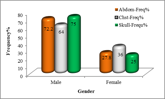 Figure 1: shows distribution of requested radiographic based on gender
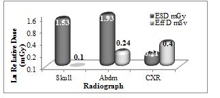
Figure 2: shows the distribution of requested radiographic cases based on causes

Figure 3: shows the average ESD in mGy & EffD in mSv received by common anatomical site radiography
Recent Publications
1. Kristin B Lysdah and BjÙ‘rn M Hofmann. (2009). What causes increasing and unnecessary use of radiological investigations? A survey of radiologists' perceptions. BMC Health Services Research, 9:155. doi: 10.1186/1472-6963-9-155.
2. United Nations Scientific Committee on the Effects of Atomic Radiation. (2000). Sources and effects of ionizing radiation: report to the General Assembly, annex D, medical radiation
3. ICRP. (1991). Recommendations of the ICRP publication 60, Annals of ICRP, Pergamon Press, Oxford.
Abdulrahman A. S. Alsayyari
Qassim University, Saudi Arabia
Title: Study of common requested radiographs and relative exposure dose in Qassim province

Biography:
Abdulrahman Alsayyari, is a vice dean of the college of applied medical sciences at Qassim university. He obtained his PhD degree from university of Queensland at Australia. He worked in both sector clinical and educational where he developed his experience and knowledge to improve the healthcare services for the Saudi population.
Abstract:
The objective of the article was to study the common requested radiographs and relative exposure dose in Qassim province in Kingdom of Saudi Arabia. The method was retrospective and analytical study for collected variables as radiographs, relative entrance surface dose (ESD) and the effective dose, patient age, gender and causative factors. The doses have been derived from the product of system output, mAs, back scatter factor BSF, focal detector distance FDD and focus – skin- distance FSD based on the equation stated by ICRU, (2005) and Davies et al, (1997):
Dose (mGy) = (Output(mGy⁄mAs)×(mAs)×(BSF)×〖(FDD)〗^2)/〖(FSD)〗^2
Then the effective dose in mSv has been derived from the equation stated by ICRP, 2007 report 103.
EffD= ∑(W_T [H_T (female)+ H_T (male)])/2
Where WT refers to weighting factor for organ or tissue and HT refers to equivalent dose to organ or tissue.
The analysis with excel software revealed that: the common requested radiographs were skull, abdomen and chest with male incidence as 75%, 72.2% and 64% respectively relative to whole sample. Traffic accident (71%) and fall-down (45%) were the most causative factors among male, female respectively, with injuries as skull fissure fracture (77%), and intracranial hemorrhage (23%). The skull radiographs noted among the age group of 11-21 years and peaking at 36% among the age group of 22-32 years. The requested abdominal radiographs appeared among the age group of 13-21 years; with frequency of (19%) and peaking at 30% among the age group of 22-30 years; with injuries as spleen ruptures (42%) and liver (27%). The chest radiographs observed among age group of 3-13 years; with frequency of 4% and peaking among age groups of 14-24 & 25-35 years old with frequencies of 19% and 21% respectively, and injuries as ribs fracture (55%), ribs dislocation (15%), pierced lung (20%) hemorrhage (10%). The average ESD for abdomen, skull and the chest radiographs were 1.93±0.8, 1.53±0.6 and 0.21±0.2 mGy, which were increase linearly following the aging, and the average effective doses were 0.24±0.1, 0.1±0.1 and 0.4±0.2 mSv respectively.
Image :
|
https://d2cax41o7ahm5l.cloudfront.net/cs/upload-images/imaging2017-33417.png |
Figure 1: shows distribution of requested radiographic based on gender
|
https://d2cax41o7ahm5l.cloudfront.net/cs/upload-images/imaging2017-17136.JPG |
Figure 2: shows the distribution of requested radiographic cases based on causes
|
https://d2cax41o7ahm5l.cloudfront.net/cs/upload-images/imaging2017-17136.JPG |
Figure 3: shows the average ESD in mGy & EffD in mSv received by common anatomical site radiography
Recent Publications
1. Kristin B Lysdah and BjÙ‘rn M Hofmann. (2009). What causes increasing and unnecessary use of radiological investigations? A survey of radiologists' perceptions. BMC Health Services Research, 9:155. doi: 10.1186/1472-6963-9-155.
2. United Nations Scientific Committee on the Effects of Atomic Radiation. (2000). Sources and effects of ionizing radiation: report to the General Assembly, annex D, medical radiation
3. ICRP. (1991). Recommendations of the ICRP publication 60, Annals of ICRP, Pergamon Press, Oxford.
Masoud Hashemi
Shahid Beheshti University of Medical Sciences, Iran
Title: Ultrasound guidance for interventional pain management of lumbar facet joint pain: An anatomical and clinical study

Biography:
Abstract:
Hissa Mohammed
National center for Cancer Care and Research, Qatar
Title: The efficiency of good communication between radiographer and autism pediatric patient in reduction of radiation dose to patient
Biography:
I completed my three-year diploma in medical radiography from Health Science School–College of North Atlantic–Qatar in 2008. Subsequently, I worked as a radiology technologist for Hamad Medical Corporation, Qatar, for two years. In 2010, I went to Edinburgh to continue my studies and obtain a bachelor’s in medical radiography from Queen Margaret University,Edinburgh. In 2014, I completed my bachelor’s and returned to Qatar, where I worked as a technologist. In 2016, I was promoted to technical supervisor at the National Center of Cancer Care and Research. I am always taking an active part in improving and developing my imaging skills, especially in pediatric imaging. In turn, I am sharing this knowledge as I train my colleagues and new staff in the department.
Abstract:
Chih-Jen Hung
Taichung Veterans General Hospital, Taiwan
Title: MR myelography for postoperative orthostatic headache
Biography:
Abstract:
We report the case of a man who presented with orthostatic headache postoperatively. The procedure of mid-thoracic epidural catheterization for posthoracotomy pain management was performed before induction of anesthesia. Unfortunately, theprocedure was abandoned due to some fluid aspirated from the epidural catheter. After operation, the patient complained of a severe headache for the first time when he was in the upright position. However, the findings of magnetic resonance (MR) myelography were characteristic of spontaneous intracranial hypotension (SIH), and the patient was successfully treated by an epidural blood patch at T1 level. Some reports implied that epidural anesthesia/analgesia might be a possible triggering factor of SIH in patients with an underlying structural weakness of spinal meninges. MR myelography can detect the pooling of CSF leakage. The T2-weighed MR image of our patient revealed dural sinus engorgement with contrast enhancement along bilateral nerve root sleeves of 5 levels from C7-T1 toT5-6 and left nerve root sleeve of C6-7 which indicated the presence of multiple simultaneous leaks at different spinal levels. The present case suggests that MR myelography could be carried out in patients with postoperative orthostatic headache when accidental dural puncture was not confirmed.
Image
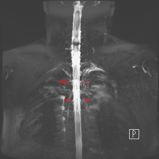
Figure 1: T2-weighed MRI revealed contrast enhancement along neural sleeves of C7-T1.
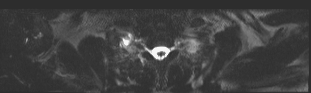
Figure 2: Many CSF leak lesions along multiple nerve root sleeves.
- Medical Imaging | Radiology Trends and Technology | X-Ray and PET
Location: Sunset -1
Chair
M. A. Alnafea
King Saud University, Saudi Arabia
Session Introduction
M. A. Alnafea
King Saud University, Saudi Arabia
Title: Non-monte carlo methods for investigating the application of coded aperture breast tumour imaging
Biography:
Abstract:
Monica Kansal
Jaypee Hospital, India
Title: Dynamics of folliculogenesis – sonographic assessment and applications in infertility management
Biography:
Monica Kansal has been practising Radiology at eminent hospital and educational institutions from last nine years with special interest in Women’s imaging.
Abstract:
Trans-vaginal sonography along with colour Doppler is the gold standard investigation in assessment of gynaecological and reproductive disorders in females. Besides exclusion of uterine, endometrial and tubal causes, sonography provides noninvasive tool for monitoring individual follicles during menstrual cycle and response to ovarian stimulation. This paper describes various uses of ultrasound in assisted reproductive techniques as the principal non-invasive modality for evaluation of key process of ovarian function – the process of folliculogenesis. Folliculogenesis refers to all phases that a primordial germ cell should pass to become mature healthy oocyte that is subsequently fertilized. It is a constant process that starts in embryogenic period and ends with the disappearance of last functional follicle in the period of menopause. Recognising the quality of follicle, its growth pattern and vascularity has a prognostic value for outcome of assisted reproduction techniques.
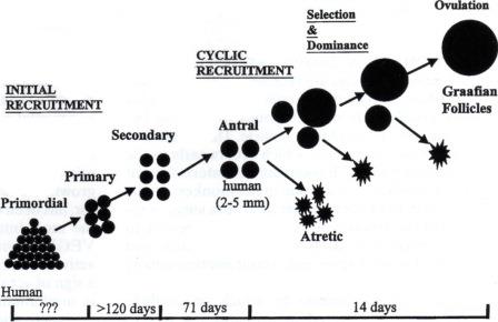
Figure 1: Folliculogenesis - Process of follicle recruitment and development.
Recent Publications
1. V Vlaisavljevic and M Dosen (2007) Clinical applications of ultrasound in assessment of follicle development and growth. Donald School Journal of Ultrasound in Obstetrics and Gynaecology 1(2):50–63.
2. Nanette Santoro, Barbara Isaac, Genevieve Neal – Perry, Tovaghol Adel, Laura Weingart and Aimee Nussbaum (2011) Impaired folliculogenesis and ovulation in older reproductive aged women. Journal of clinical endocrinology and metabolism: 88(11):5502–5509.
3. Angela Baerwald, Gregg P Adams and Roger A Pierson (2012) Ovarian antral folliculogenesis during human menstrual cycle: A review. Human Reproduction Update, 18(1):73-91.
David Sipos
University of Pecs Faculty of Health Sciences, Hungary
Title: Radiographer prospects in Hungary
Biography:
Abstract:
Vikas Leelavati Balasaheb Jadhav
Dr.D.Y.Patil University, India
Title: TransAbdominal sonography of the small & large intestines

Biography:
Abstract:
Amjed Eljaili
Ysbyty Gwynedd, UK