Day 2 :
Keynote Forum
Daniel L. Farkas
University of Southern California, USA
Keynote: Multimodality Imaging for clinical research: The role of biophotonics
Time : 10:00-10:50

Biography:
Daniel L Farkas is a former Fulbright scholar, directed a National Science and Technology Center at Carnegie Mellon University. He was Professor of Bioengineering at
University of Pittsburgh, and Vice-chairman for research and; Professor of Surgery at Cedars-Sinai Medical Center. He has 200+ publications, 11 editorial boards, $80
million funding, 32 international conferences chaired, and several prestigious awards. His focus is on translational biomedical optics, in academia and startups he founded.
Abstract:
In order to see the bench-to-bedside dream of translational research become a reality, we need to develop imaging approaches
that is technologically sophisticated, allow deployment into a clinical setting. Multimodality imaging is gaining adoption, but
the bio-photonic component is absent or un-sophisticated. Our focus area is where light and patient meet, and improvements
that yield better outcomes, by identifying/addressing the obstacles preventing timely clinical adoption of laboratory-based
advances, not the least of which is the difficulty of detecting, characterizing and monitoring very small entities (molecules,
cells) within the human body, especially quantitatively, dynamically, and preferably without contrast agents. How and where
we look becomes critically important, especially if one targets (as one should) early diagnosis; for this, new tools and strategies
are needed, with likely new outcomes. We proposed and implemented an optical multimode approach to biomedical optical
imaging at all levels, featuring hyperspectral imaging, and optimized for earlier, more quantitative/reproducible detection of
abnormalities and a tighter spatio-temporal coupling between such diagnosis and intervention. Addressing major areas of
unmet need in the clinical realm with these new approaches should yield important improvements in disease management.
Our work on cancer, stem cells, vascular and neuro (specifically highlighting very early detection of Alzheimer’s disease)
applications will be described, with emphasis on the new technologies needed to achieve the desired imaging performance.
Thoughts about better ways for academia, the clinical and the corporate world to work together for innovative imaging solutions
and their use for addressing major disease will be briefly outlined.
- Magnetic Resonance Imaging | Ultrasound | Clinical Research
Location: Sunset -1

Chair
Daniel L. Farkas
University of Southern California, USA
Session Introduction
Hissa Mohammed
National Center for Cancer Care and Research, Qatar
Title: The efficiency of applying the radiology technologist of the Radiation dose monitoring technique during the fluoroscopy procedures for oncology pediatric aged between 4 – 7 years old
Biography:
Hissa Nasser Mohammed completed his Diploma in Medical Radiography from Health Science School, College of North Atlantic, Qatar in 2008. He worked as
Radiology Technologist at Hamad Medical Corporation, Qatar for two years. In 2014, he completed his Bachelor degree in Medical Radiography from Queen
Margaret University, Edinburgh. He was Technical Supervisor at National Center of Cancer Care and Research.
Abstract:
Fluoroscopy is one of the radiation sources used in diagnostic processes in radiology. Owing to the diagnostic approach that entails observation of the affected anatomy using radiation in real time, harmful effects may potentially occur. Therefore, safety measures in the radiographer’s use of equipment and effective monitoring and management are essential in diagnostic processes. Additionally, patients in the age group of four to seven years have less anatomy and tissue development, which presents higher levels of health risks. The study will focus on analyzing the pre-procedure requirements and the set of guidelines, which enable the enforcement of the safety of the concerned patients within the pediatric practice. For example, the study will observe the potency of undertaking the reduction of the fluoroscopic times and improving the communications between the health specialists. The study will also evaluate methods and techniques employed in achieving efficiency in fluoroscopy radiation dose management. Direct and indirect methods will be employed to monitor the dosage effects. Direct methods would entail performing a skin dose test on the target area of fluoroscopic radiation. Detectors would be employed, for example the photographic films and thermoluminescent dosimeters. The indirect methods will employ the use of the dose area product meter to ascertain the effects of radiation on the patients. Some of the dose reduction techniques involve the manipulation of equipment operation, for example beam quality adjustment, dose level setting, and dose spreading. The results used to evaluate the dosage level will entail analyzing measurements of the skin exposure unit of the fluoroscopic equipment. Additionally, results from the change in beam quality and the effect on the skin will be analyzed in the study. These results will be achieved through the use of different operational voltage levels on the fluoroscopic equipment.
Skin dose will be determined through a combination of several measurable factors in fluoroscopic equipment operation. Due to the wide adoption of the fluoroscopic radiation technique in the pediatric oncology diagnostic process, dose monitoring is important in ensuring patient safety. Direct monitoring such as the skin dose procedure is effective, due to the reduced risk, which would otherwise result from other methods. Notable measures for efficiency in dose monitoring require adequate training of fluoroscopic operators on appropriate equipment use and continual observance of quality control procedures. These interventions and procedures should not compromise the quality of imaging and dose specification.
Daniel L. Farkas
University of Southern California, USA
Title: High resolution, non-invasive multimode optical imaging: A proposed diagnostic and assessment tool in Alzheimer’s Disease

Biography:
Daniel L Farkas is a former Fulbright scholar, directed a National Science & Technology Center at Carnegie Mellon University. He was Professor of Bioengineering at University of Pittsburgh, and Vice-chairman for research and Professor of Surgery at Cedars-Sinai Medical Center. His scientific interests center on investigating the living state with light, for uses in biology, bioengineering, medicine and surgery. He published 200+ articles and 28 books. He chaired 30+ international conferences.
Abstract:
Statement of the Problem: Alzheimer’s disease (AD) is a major unmet health challenge characterized by fast increasing incidence and costs.
Methodology: Our group has recently introduced optical imaging in the retina as a non-invasive method for mapping the occurrencesize and location of beta amyloid plaques, the primary pathology in AD. We have shown that using the fluorescence of curcumin,which attaches specifically to these plaques, we could quantitate the features of these plaques, including in vivo, and even document their reduction by immune treatments. These preclinical studies were also extended to the clinical domain, by using archival human eyes from patients with known levels of AD, as assessed both by brain histopathology and cognitive impairment (prior to death). We present a method for extending such studies to living patients, still using the retina as the window to the brain and plaques as indicators, but without the use of an extrinsic biomarker such as curcumin. This raises the level of experimental difficulty, thus requiring new technologies that we invented.
Findings: We designed a multimode optical imaging instrument, essentially a new type of confocal scanning laser ophthalmoscope,with some (needed) performance advantages over current commercial offerings. Our system consists of the following elements, all proprietary, and patent-protected: A highly versatile light source: pulsed, 400-1400 nm, with ~1 nm resolution; a new galvanometric method of scanning, with synthesized pivot point, not requiring a custom coupling lens; spectral analysis of imaging data, including
hyperspectral image segmentation and elimination of background; a more sensitive method of detecting light via parametric.
Conclusion & Significance: This new instrument achieves significant improvements in all of the following: spatial resolution,imaging depth, imaging angle in the retina (and thus spatial coverage), sensitivity and specificity. It will be used to image, fast and non-invasively, amyloid plaques in the retina, and any other retinal features of interest. We envisage that this instrument and the
approach it enables should be used in AD drug/treatment trials, as it allows the repeatable, non-invasive and quantitative imaging of amyloid plaques (via both their autofluorescence and scattering), and of their relationships with important structures in the eye, such as blood vessels.
Claudia Paola Rivera-Uribe
Nuevo León Autonomous University, Mexico
Title: Diaphragmatic shortening fraction and pulmonary ultrasound combined analysis for extubation success prediction in critical care patients
Biography:
Claudia Paola Rivera Uribe has completed her medical school at the age of 24 years from Guadalajara University and posgrade studies from Nuevo León
Autonomous University. She is Chief of Residents of Pulmonary and Critical Care Medicine.
Abstract:
Invasive respiratory support is a cornerstone of Critical Care Medicine, however, protocols for withdrawal of mechanical ventilation are still far from perfect. Failure to extubation occurs in up to 20% of patients, despite a successful spontaneous breathing trial (SBT).We prospectively included ventilated patients admitted to medical and surgical intensive care unit in a university hospital in northern Mexico. At the end of a successful SBT, we measured Diaphragmatic Shortening Fraction (DSF) at the end of inspiration and at the end of expiration, and the presence of B-lines in five zones of the right and left lung. The primary objective was to determine whether analysis of DSF and Pulmonary Ultrasound improves prediction of extubation success. Eighty-two patients were included,24 (29.2%) failed to extubation. At univariate analysis, DSF (Youden’s J: > 30% [sensibility and specificity 62 and 50%, respectively])and number of B-lines zones (Youden’s J: > 1 zone [sensibility and specificity 66 and 92%, respectively]) were significant related to extubation failure (area under the curve 0.664 [0.526 to 0.801] and 0.819 [0.703 to 0.934], respectively). At the binomial logistic regression, only the number of B-lines zones remains significantly related to extubation failure (OR 5.91 [2.33-14.98], p < 0.001). In patients with a successfully SBT, the absence of B-lines significantly decreases the probability of extubation failure. DSF analysis does not add predictive power over the use of pulmonary ultrasound.
Ala khasawneh
University of Pécs, Hungary
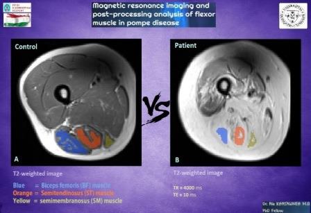
Title: Magnetic resonance imaging and post-processing analysis of flexor muscle in lateonset Pompe disease

Biography:
Ala Khasawneh is a Jordanian Doctor. He completed his Diploma in General Medicine (MD) and has been awarded the qualification of a Physician and title of
Doctor of Medicine from National Pirogov Memorial Medical University. He worked in Basma Hospital, Jordan. Currently, he is a PhD fellow in Diagnostic Medical Imaging in Hungary. As a Doctor, his main interest is to create new pathways for improving health care.
Abstract:
Pompe disease is a rare multisystem genetic disorder that characterized by a deficiency of the lysosomal enzyme acid alphaglucosidase and cytoplasmic glycogen accumulation causing damage that leads to muscle weakness. This study aim is to evaluate the muscle MRI pattern of twelve adults with late onset Pompe disease and twelve sex- and age-matched healthy controls (Age
range 19-59) for feature extraction which will be used to identify and classify functioning and non-functioning muscles. A training procedure was implemented using an Exercise Dynamometer device to stimulate the muscle for maximal contraction, MRI images data was used to compare the three flexor muscles in the lower limb function. MRI images data of biceps femoris (BF) muscle,Semitendinosus (ST) muscle, semimembranosus (SM) muscle before and after exercise (base, 30 min, 24 hours) was measured.
We performed and quantified T2 relaxation data of flexor muscles, and all data analyzed using repeated measure ANOVA to compare within related groups of the independent variable time (Base, 30M, 24H). According to our results, the significantly lower T2 value in the ST muscle of controls was observed (base=43ms, 30min=48ms, 24h=43ms; P < 0.05), but the change in SM muscle and BF muscle were not significant. In patients, we detected significantly higher T2 value in SM muscle evolve over time (base=129ms,30min=132ms, 24h=128ms; P < 0.05) compared to the controls, but ST muscle neither BF muscle doesn't show significant change.
As a conclusion, we can say that in Pompe patients the SM muscle can only react to the exercise apparently and shows us an activity in affected muscle cells, compared to the BF and ST muscle not shown any activity, that’s mean perhaps the Pompe disease change the muscle cells structure to interact to the exercise.
Syed Muhammad Anwar
University of Engineering and Technology, Pakistan
Title: Deep learning in medical image analysis
Biography:
Syed Muhammad Anwar is assistant professor at department of Software Engineering, University of Engineering, and Technology, Taxila and leading the Signal,image and multimedia, processing, and learning (SIMPLe) group. His research interest includes magnetic resonance imaging, machine learning, deep learning,medical image analysis and wearable and m-health.
Abstract:
Medical image analysis is the science of analysing or solving medical problems using different image analysis techniques for affective and efficient extraction of information. It has emerged as one of the top research area in the field of engineering and medicine. Recent years have witnessed rapid use of machine learning algorithms in medical image analysis.
These machine learning techniques are used to extract compact information for improved performance of medical image analysis system, when compared to the traditional methods that use extraction of handcrafted features. Deep learning is a breakthrough in machine learning techniques that has overwhelmed the field of pattern
recognition and computer vision research by providing state-of-the-art results. Deep learning provides different machine learning algorithms that model high level data abstractions and do not rely on handcrafted features.
Recently, deep learning methods utilizing deep convolutional neural networks have been applied to medical image analysis providing promising results. The application area covers the whole spectrum of medical image analysis including detection, segmentation,classification, and computer aided diagnosis. A brief introduction to the application of deep learning algorithms in medical image retrieval, segmentation, and detection will be presented.
Kunwarpal singh
Sri Guru Ram Das Institute of Medical Sciences and Research, India
Title: Role of conventional and diffusion weighted MRI with ADC values in staging of carcinoma of prostate and response to treatment wherever possible with clinical and biochemical correlation
Biography:
Kunwarpal Singh is Associate Professor in Department of Radiodiagnosis at Sri Guru Ram Das Institute Of Medical Sciences And Research, Punjab. He gave 13
publication and presentations in National and International Journals and conferences.
Abstract:
Background & Aim: Prostate cancer is one of the commonest malignancies in men that cause significant morbidity and mortality worldwide. The latest population-based cancer registry in India by ICMR records the age adjusted rate (AAR) to be 8.4, 10.7, 7.7,and 1.9 per 100,000 populations in the cities of Bangalore, Delhi, Mumbai and rural Barshi respectively. MRI is a powerful imaging modality for evaluation of anatomy and pathology of the prostate. This study aimed to evaluate the role of conventional MRI sequences
with diffusion weighted imaging using ADC values in staging of prostate carcinoma and in assessing response to treatment.
Materials & Methods: We prospectively studied 75 patients who came with suspicion of having carcinoma of prostate on Philips Gyroscan Achieva 1.5 Tesla MRI using sense body coil. Morphologic features of lesions were assessed on conventional sequences as well as on diffusion weighted imaging in pretreated and post treated patients. ADC values were calculated and statistically analysed.
Results: Highly significant correlation was found between serum PSA levels, prostate size, and staging and ADC values in both pretreated and post treated patients with p value <0.05. However, conventional weighted imaging did not show any significant change in signal intensity in post treated patients suggestive of added advantage of DWI.
Conclusions: MRI is a powerful imaging modality for comprehensive structural evaluation of anatomy and pathology of prostate
gland. Addition of ADC and DWI to T2WI provide more accurate results for prostate cancer detection, staging and post treatment
response.
Shajeem Shahudeen
Vivid Diagnostic Centre, India
Title: Role of CT in screening coronary artery disease
Biography:
Shajeem Shahudeen has completed his MD in Radio Diagnosis from DY Patil University, Navi-Mumbai. He is the Managing Director and Consultant Radiologist at Vivid Diagnostic Centre, Kochi, India. He has also done several papers, poster presentation and publications.
Abstract:
Coronary artery disease (CAD) remains the leading cause of death in western countries with increasing prevalence in developing countries. The standard reference for diagnosis of CAD is coronary catheter angiography. Imaging of the heart has always been technically challenging because of the heart’s continuous motion.
CT imaging of the heart moved into the diagnostic realm by the introduction of multi-detector row CT (MDCT) and development of ECG-Synchronized scanning and reconstruction techniques.
These modalities allow for faster volume coverage, high spatial and temporal resolution. The introduction of MDCT especially has greatly benefitted cardiovascular CT applications as the speed of image acquisition shortens, breath hold and examination time for the patient and reduces the amount of contrast media needed for high and consistent vascular enhancement. The advents of 128-slice MDCT scanner sub millimetre resolution (0.4 mm) of substantial anatomic volumes are routinely achieved.
Aim of this study is: to study the role of MDCT coronary angiography in coronary artery disease (CAD) in symptomatic and asymptomatic patients; to study the calcium score in patients undergoing MDCT coronary angiography; to study the role of MDCT coronary angiography in patients with risk factors and to study the role of MDCT coronary angiography in follow ups of post-CABG and post-angioplasty stent patients.
Dongyeon Lee
Yonsei University, South Korea
Title: Reconstruction quality in 4D cone-beam computed tomography (CBCT) incorporated with deformable image registration (DIR) method
Biography:
Dongyeon Lee completed his MS degree from Yonsei University, Republic of Korea, and is a PhD candidate in the Department of Radiation Convergence
Engineering at Yonsei University. He has published more than 10 papers in reputed journals and his research interests include CT, image registration, GPU
acceleration, and so on.
Abstract:
In conventional three-dimentional (3D) cone-beam computed tomography (CBCT) reconstruction, the image quality of reconstructed images is typically degraded due to patient’s motion such as respiration and heartbeat. In order to solve these difficulties, 4D CBCT incoporated with a phase-angle sorting scheme is often utilized. However, in this case, it requires dense projections in each phase to obtain reconstructed images of high quality and also severe aliasing artifacts are often present due to the limited number of projections at each phase used in the reconstruction. In this study, as an alternative, we propose an effective method for reducing motion blur and aliasing artifacts in conventional 4D CBCT reconstruction, the so-called deformable image registration(DIR), and made a quantitative comparison of the reconstruction qualities by both shcemes. In the proposed method, DIR process is carried out in two different ways as follows. In the first way, DIR process is applied to the resultant reconstructed images in 4DCBCT, while in the second way to the original projections on the basis of the reconstructed projections that are produced by forwardprojecting the reconstructed CBCT images in a single phase. The latter process is possibly performed using the adaptive steepestdescent POCS (ASD-POCS) algorithm for high reconstruction quality. Our results indicate that the motion blur and aliasing artifacts in the reconstructed CBCT images by using the DIR method were more significantly reduced, compared to the phase-angle sorting method. In addition, the second method in the DIR process seems more effective for reducing imaging dose than the first method.