Day 1 :
Keynote Forum
Ken Miles
University College London, UK
Keynote: Immune checkpoint blockade for solid tumors: Opportunities for imaging to contribute to precision medicine

Biography:
Ken Miles is Honorary Professor at the Institute of Nuclear Medicine, University College London and Senior Medical Officer at the Princess Alexandra Hospital, Brisbane. He is dual-trained in Radiology and Nuclear Medicine and has wide experience in Positron Emission Tomography, having been involved in the establishment of four PET centres worldwide. His research interests include the development and evaluation of advanced quatitative imaging techniques in oncology. He has authored or co-authored more than 130 peer-reviewed scientific publications and contributed to or edited 11 books in Radiology. Ken is a past editor in chief of the journal Cancer Imaging.
Abstract:
Overall tumour mutation burden within solid tumours may be predictive for clinical response to immune checkpoint blockade but can be difficult to assess within routine clinical practice. This presentation discusses the potential for an alternative image-based approach to patient selection. Neoantigenic diversity associated with a high mutation burden results in tumour-associated inflammation. A potential imaging correlate for this process is provided by dual time point PET (DTP) with fluorodeoxyglucose (FDG). In comparison to tumour, inflammatory cells show reduced retention of FDG at later time points due to expression of glucose-6-phosphatatse. Low FDG retention on DTP has been shown to have prognostic significance for several tumour types and initial data indicates reduced FDG retention in metastatic lesions from tumour types known to exhibit high mutation rates. The evolutionary dynamics of malignancies with high mutation burdens typically results in the co-existence of genetically distinct sub-clones within the same tumour. Imaging methods such as CT texture analysis (CTTA) that quantify macroscopic tumour heterogeneity can potentially provide a correlate for this clonal development. In NSCLC, CTTA has been shown to be prognostic for a range of tumours and can demonstrate differences in heterogeneity between wild-type tumours and those exhibiting driver mutations known to be associated lower or greater mutation burden. Radiogenomic approaches that seek to identify imaging correlates for tumour mutation burden warrant further exploration as non-invasive and potentially cost-effective means to select patients for immune checkpoint blockade.
Keynote Forum
Tanya Moseley
University of Texas MD Anderson Cancer Centre, USA
Keynote: Impact on clinical management of after-hours emergent or urgent breast ultrasonography in patients with clinically suspected breast abscesses
Time : 10:15-10:55

Biography:
Tanya W. Moseley, MD has distinguished herself as a top-notch radiologist, clinician, educator, researcher, and leader in her field. She is a world-class teacher of undergraduates, residents, fellows, medical students, and breast imaging technologists having supervised and trained numerous visiting scientists, residents, and fellows over the past 20 years. She received the 2017 University of Texas Regents’ Outstanding Teaching Award. Dr. Moseley is a former Fellowship Director of Breast Imaging, and developed an outstanding Breast Ultrasound Course at MD Anderson Cancer Center in Houston, Texas. She is the past Breast Section Program Chair and Breast Section Course Director of the American Roentgen Ray Society (ARRS) Case-Based Imaging Review Breast Section. Dr. Moseley received her Doctorate of Medicine with Honors at the University of Iowa College of Medicine in Iowa City, Iowa. She entered a Clinical Residency in Diagnostic Radiology at the Mayo Clinic Graduate School of Medicine in Rochester, Minnesota, and continued on at the Mayo Clinic in a Clinical Fellowship in Mammography and Thoracic Imaging. After completing her fellowship, Dr. Moseley joined Mayo Clinic as a Senior Associate Consultant, and then joined the Division of Diagnostic Imaging at MD Anderson Cancer Center. She is presently a Professor of Diagnostic Radiology and Breast Surgical Oncology at MD Anderson.
Abstract:
Newly diagnosed breast abscesses are generally treated as a medical emergency that may necessitate immediate interventional treatment. At our institution, there is no in-house after-hours coverage for breast ultrasonography. We could find no peer-reviewed studies on the cost-effectiveness or clinical management impact of on-call ultrasound technologist coverage for imaging of breast abscesses. The purposes of this study were to determine the incidence of breast abscess in patients with clinical findings highly suggestive of abscess, identify clinical factors associated with breast abscess in such patients, and determine the impact of after-hours emergent or urgent breast ultrasonography on the clinical management of breast abscesses in both outpatients and inpatients. We retrospectively reviewed 100 after-hours breast ultrasound studies performed at our tertiary care center from 2011 to 2015 for evaluation of a suspected breast abscess. Only 26% of our patients with clinically suspected abscess ultimately had a confirmed abscess. Factors associated with breast abscess were a palpable abnormality and a history of breast surgery within the eight weeks before presentation. After-hours diagnosis of an abscess was associated with after-hours clinical intervention. Of the 74 patients in whom after-hours ultrasound imaging showed no evidence of abscess, only three patients underwent after-hours drainage. Our findings support overnight and weekend breast ultrasound coverage in large tertiary care centers.
Keynote Forum
Hyoung K Lee
Missouri University of Science and Technology, USA
Keynote: Advances in diagnostic X-ray sources

Biography:
Dr. Hyoung Lee has his expertise in radiation detection and imaging. He has worked in teaching hospitals as a medical physicist and has also been a faculty member in academia since receiving his PhD. His research area includes radiation detector design and characterization, advanced radiation sources, advanced imaging systems using x-ray, gamma ray or neutron, algorithms for CT reconstruction, image processing, image analysis, and application of deep learning for medical imaging. He has successfully conducted many research projects that covered a wide spectrum of radiation detection and imaging technologies, from development of radiation detectors to development of image enhancement algorithms (some of which were transferred to industry). He also helped a medical imaging company develop flat-panel x-ray image sensors, digital radiography systems, digital dental imaging systems and cone-beam CT systems. His current research includes development of flat-panel x-ray sources, compact x-ray tubes for stationary cardiac CT and switchable radioisotopes.
Abstract:
X-ray imaging has been a prominent part of diagnostics since x-ray was discovered by Röntgen in 1895. Over the past ~120 years, the x-ray source technology has undergone noteworthy advancements that resulted in a wide range of x-ray imaging modalities for diagnosing various ailments in human anatomy. With the contribution of researchers from all over the globe, the x-ray source technology has been continually advanced to overcome the technical challenges behind applying the x-ray imaging technology to various imaging modalities. In this talk, the advances in diagnostic x-ray sources and their applications that have already being implemented in medical practice and that have a potential to make it to medical practice in the near future will be discussed. It will start with a brief history of x-ray sources along with major leaps in technology that overcame challenges such as dissipating large amount of heat from the anode, maintaining vacuum pressure in the tube, extending the tungsten filament lifetime, and avoiding high-voltage breakdown. Other technical challenges and issues of certain x-ray imaging systems, for example, cardiac CT, duel-energy (spectral) CT, etc. will be discussed. Recent research activities in x-ray tube technology such as utilization of field emission electron source in place of filament, transmission type anodes, distributed x-ray sources, and flat-panel x-ray sources (Fig. 1) will be introduced. In addition, advanced diagnostic imaging systems such as stationary CT and tetrahedron beam CT based on advanced architecture of x-ray tubes will be introduced. The stationary CT system implements a stationary gantry with x-ray sources and detectors distributed along the gantry to electronically sweep an x-ray beam along the gantry (Fig. 2), which is expected to reach a temporal resolution of ~30 msec. Finally, nonbremsstrahlung x-ray sources that may be useful for medical imaging in the future will be introduced.
Keynote Forum
Mariela Agolti
Centre of Nuclear Medicine, Argentina
Keynote: Review of our experience in 18 F-Choline PET CT in the diagnosis of bone metastasis for prostate carcinoma with biochemical relapse
Biography:
Dr Mariela Agolti is a certificated Nuclear Medicine Specialist. She serves as the Medical Director of Centro Medicina Nuclear Clinica Modelo, Parana, E Rios Argentina. She is also the Director of University Technology Career Bioingeneering University and is the member of WARMTH.
Abstract:
Purpose : To review the bibliography and our results in the usefulness of 18 F-Choline PET fused with CT in prostate cancer to diagnose bone metastasis. Prostate cancer is the most common cancer in men in our country.
Methods: 18Fluorcholine ( 18FCH) PET CT was performed in 27 patients with prostate cancer using 0,035 Mci/kg in our department of nuclear medicine since 31/ 01/2017 to 01/08/2018 , we included patients with biopsy-proven prostate cancer with rise in PSA for restaging.
In our patient series, biochemical recurrence of prostate cancer was considered present when the levels of serum PSA measured after surgery or radiotherapy and/or hormonal therapy exceeded 0.1 ng /ml . Results were correlated with clinical, bio chemical and imaging follow up.
Results: We studied 27 patients. We found bone metastases in 10 patients, 15 patients were high risk patients, considering high risk when PSA was 10 or more and Gleason was 7 or more. We found hypermethabolic bone metastasis in 9 of this patients , only 2 of them had blastic images in CT with normal 18 FCH uptake, however in 2 of the 10 patients with multiple bone metastasis , At least 50%of the lesions had no 18 FCH uptake . They were predominantly blastic.
Conclusion: 18 FCH PET CT is a very useful method for diagnosing bone metastasis in Prostate Cancer biochemical recurrence , however when they are very hyper dense in CT they may be not uptake 18 FCH , in those cases the CT was used for bone metastasis diagnostic.
Keynote Forum
Partha S Choudhury
Rajiv Gandhi Cancer Institute and Research Centre, India
Keynote: Precision management in castration resistant prostate cancer (CRPC) : A theranostic approach
Time : 12:35-13:15

Biography:
Dr Partha S Choudhury is an internationally acclaimed leading Nuclear Medicine Physician of India with special interest in Radionuclide Therapy of various types of cancers. He has more than 25 years of experience in Nuclear Oncology. He is heading the department of Nuclear Medicine in Rajiv Gandhi Cancer Institute & Research Centre Delhi India since 1998 and has been instrumental in its sustained growth over the last 20 years. He has introduced and standardized new procedures in the department both in terms of disease specific diagnostic and molecular imaging & molecular therapy. He is an invited speaker in conferences and symposiums across many countries, the main ones being United Kingdom, Austria, South Africa and South America. He is an avid clinical researcher with publications in peer reviewed journals. He is a technical co-operation consultant & participant of co-ordinated research projects of International Atomic Energy Agency (IAEA) Vienna
Abstract:
The term theranostics is the combination of a diagnostic tool that helps to define the right therapeutic tool for specific disease. It signifies the “we treat what we see & see what we treat” concept. A diagnostic radionuclide is labelled with the target and once expression is documented, the same target is labelled with a therapeutic radionuclide and treatment is executed. In Nuclear Medicine theranostics is easy to apply because of an easy switch from diagnosis to therapy with the same vector. This concept is utilized in few malignancies and prostate cancer is one of them. PSMA expression in normal and other tumor is less than prostate cancer cell. Its expression increases with higher grade and hormone resistance. We have reported excellent sensitivity and detection capability of PSMA for sub centimeter sized lymphnodes at staging . Besides evaluation of recurrence, 68Ga-PSMA PET can be utilized in advanced prostate cancer for detection of metastasis. 68Ga-PSMA also serves the basis of treatment of CRPC with 177Lu PSMA. Progression to androgen independence is the main cause of morbidity & death in these patients. Based on the theranostic concept the aims of treatment with 177Lu PSMA are to improve outcome by early interventions in suboptimal responders, sparing low risk patients from over treatment, reduce treatment related side effects, ensure effective palliation & improve quality of life. Tumor targeting with 177Lu PSMA saves normal tissue & delivers high dose to tumor. Easy radiopharmaceutical labelling & high expression in all cancer cells makes it an optimal target for radionuclide therapy. It is safe with a low toxicity profile. In our experience at RGCI & RC (our institute) we have seen objective regression in lesions and symptomatic relief & found it to be a safe & effective method for treating end stage androgen independent, progressive CRPC. We believe that treatment of recurrent prostate cancer is feasible with 177Lu PSMA with a positive objective response & a low side effect profile where achievable tumor dose is demonstrated by Ga-68 PSMA scan before therapy.
- Computed Tomography | Musculoskeletal Imaging | Neuroradiology
Location: Atlantis 2

Chair
Hyoung K Lee
Missouri University of Science and Technology, USA
Session Introduction
Hyoung K Lee
Missouri University of Science and Technology, USA
Title: Compact X-ray tubes for stationary CT architecture

Biography:
Dr. Hyoung Lee has his expertise in radiation detection and imaging. He has worked in teaching hospitals as a medical physicist and has also been a faculty member in academia since receiving his PhD. His research area includes radiation detector design and characterization, advanced radiation sources, advanced imaging systems using x-ray, gamma ray or neutron, algorithms for CT reconstruction, image processing, image analysis, and application of deep learning for medical imaging. He has successfully conducted many research projects that covered a wide spectrum of radiation detection and imaging technologies, from development of radiation detectors to development of image enhancement algorithms (some of which were transferred to industry). He also helped a medical imaging company develop flat-panel x-ray image sensors, digital radiography systems, digital dental imaging systems and cone-beam CT systems. His current research includes development of flat-panel x-ray sources, compact x-ray tubes for stationary cardiac CT and switchable radioisotopes.
Abstract:
For imaging arrhythmia patients with cardiac computed tomography (CT), a temporal resolution <50 msec is desired, which cannot be delivered by conventional CT architectures with a rotation gantry. By eliminating the physical rotation of the gantry and electronically sweeping x-ray beams across the gantry, stationary CT architecture can achieve a temporal resolution <50 msec. Stationary CT architecture utilizes a stationary gantry comprising separate arrays for distributed x-ray sources and detectors (Fig. 1). These individual x-ray sources are required to be compact, fast, and individually addressable in order to acquire 200+ projections for successful CT reconstruction and achieve a temporal resolution <50 msec. A compact x-ray tube that fits these requirements is being developed.
The aim of the present research was to develop and study the first-generation prototype compact x-ray tube primarily consisting of a CNT-based cold cathode and a transmission type anode (Fig. 2). Monte Carlo simulations were conducted to compare tungsten, molybdenum, and rhodium as target materials and to optimize the target thickness for a transmission type anode. Using a particle-in-cell technique, the electron focusing was studied to design an electrostatic focusing lens for achieving <1 mm focal spot size (FSS) of x-ray generation in the prototype. The prototype was studied experimentally to understand the performance of the prototype and its control parameters. The results of these simulations and experiments showed the following: CNT-based cold cathode can generate electron pulses with frequencies up to hundreds of kilohertz; an electrostatic lens can achieve the required <1 mm FSS; the lens aperture, thickness, and location can be used as a coarse control on the FSS, while the lens potential can be used as a fine control; and x-ray energy spectra from a transmission type anode is similar in shape to the energy spectra from a conventional reflection type anode after filtration.
Juan de dios Berna Mestre
University of Murcia, Spain
Title: Superficial soft tissue tumors: Role of sonoelastography

Biography:
Prof. Juan de Dios Berna Mestre has more than 10 years of hospital care as radiologist, as well as teaching at the medical school. In his research career there are publications on various topics, especially on new techniques of ultrasound. Although is main field is musculoskeletal radiology, one of its main lines of research is the development of a new technique for the imaging diagnosis of the male urethra: The Clamp Method.
Abstract:
Statement of the Problem: Sonoelastography (SE) is a new ultrasound technique that is increasingly used for the evaluation and characterisation of soft tissue lesions. Two main SE techniques are distinguished: compression sonoelastography (CSE) and shear-wave elastography (highlighting the ARFI method: Acoustic Radiation Force Impulse). The relevance of SE in the diagnosis of prostate, breast, thyroid and lymph node lesions has been reported but there are no concluding references to musculoskeletal tumors. Sometimes it is a diagnostic challenge to define the benignity or malignancy of superficial soft tissue tumors (SSTT), as well as its histological type, by means of different imaging techniques, so in most cases a biopsy is used for histological diagnosis. The purpose of this study is to describe the SE features of different histological types of SSTT. Methodology & Theoretical Orientation: During a period of 7 years, SSTT were consecutively evaluated using the CSE and ARFI techniques. An Acuson S2000 (Siemens, Germany) ultrasound machine was used. In all cases, ultrasound-guided biopsy was performed and histological diagnosis was obtained. Findings: 185 SSTT were included, of which 44.3% (n = 82) were sarcomas. The WHO classification of SSTT differences 9 histological categories and cases of all of them were obtained. In addition, other SSTT not included in this classification were included. Conclusion & Significance: The use of SE increases diagnostic accuracy in determining the histological type of SSTT.
Prashanth Balaji
Hillingdon Hospital, UK
Title: Assessment and performing CT Imaging with NICE head injury guidelines in the emergency department
Biography:
Dr Prashanth Balaji is a foundation Year 2 doctor in The Hillingdon Hospital, Uxbridge. He is interested in pursuing his future career in radiology. He has carried multiple audit and presented posters in National conference. He is an enthusiastic teacher who has organised and conducted multiple teaching sessions for medical college students and doctors . He also plays an vital role in sharing departmental responsibilities such as Rota – Coordinators, etc. He has fine tuned himself by attending multiple radiology conference and courses and looking for the 4th world congress on Medical Imaging and Clinical Research.
Abstract:
Background
Head Injury is defined as trauma of head apart face. About 1.4 million people present to ED every year. As a result, National Institute of Health & Clinical Excellence formulated clear guidelines (CG176) for accessing and performing CT Imaging. This study evaluates whether patients are accessed and performed CT Imaging according to NICE guidelines.
Methods
A retrospective study was carried out in the Emergency department with suspected head injury patients, in September 2017. Electronic clerking documentations and CT requests forms of 94 patients (64 Adults and 27 children) were analysed to check, if they were accessed for indications and performed CT Imaging according to NICE Guidelines.
Results
Among 94 patients, 23 had CT imaging of which 17(74%) met the NICE guidelines while the 6 (26%) didn’t. Results of the scans that didn’t meet criteria were normal. In addition, documentation issues were identified in clerking and CT request forms while accessing risk factors e.g. 16% and 26% of adults and children were documented for seizures respectively. Conclusions:
This retrospective study emphasises the understanding of doctors in managing Head Injury with 74% of scans compliant with Guidelines. To achieve further compliance and increase the quality of documentations, certain modifications have been implemented:
- Introduction of CT Assessment Proforma
- Presentation of findings in regional meeting
- Teaching for doctors and nurses
- Posters with key information from CG176
With these implementations, we intend to achieve 100% compliance, alongside accurate documentation ensuring imaging is carried out in a cost- and resource-effective manner while preventing unnecessary radiations.
Juan de dios Berna Mestre
University of Murcia, Spain
Title: New imaging techniques for the male urethra

Biography:
Prof. Juan de Dios Berna Mestre has more than 10 years of hospital care as radiologist, as well as teaching at the medical school. In his research career there are publications on various topics, especially on new techniques of ultrasound. Although is main field is musculoskeletal radiology, one of its main lines of research is the development of a new technique for the imaging diagnosis of the male urethra: The Clamp Method.
Abstract:
Statement of the Problem: Introduction of contrast in urethrography was first done using clamp devices coupled to a syringe. These were replaced by the conventional technique using a Foley catheter, which is the most widely used method for performing urethrography. The drawbacks of this method are that it can cause pain on inflation of the balloon and it is not useful in cases with urethromeatal alterations. Sonourethrography has been reported as being more accurate than urethrography for measuring urethral strictures and also for assessing the degree of spongiofibrosis. However, sonourethrography is not widely used, probably due to the difficulty of the technique. The purpose of this study is to describe “The Clamp Method” for performing urethrography, sonourethrography, MR and CT-urethrography. Methodology & Theoretical Orientation: The present study describes a technique to optimize the imaging diagnosis of the male urethra, using a clamp device and a fine prelubricated catheter connected to a drip infusion system. A comparative study with the conventional technique of urethrography was performed. Another study was conducted to evaluate the clamp method for sonourethrography. Findings: Urethrography could not be performed with conventional technique in the 30 percent of the cases, while all the cases were performed by clamp method. Distressing pain was reported in most cases respect inflation of the balloon, and intense pain with urethral bleeding in some of them, while no pain was reported in most cases respect external compression. Sonourethrography showed greater capacity for detecting strictures than urethrography. Conclusion & Significance: The clamp method of urethrography is simple and well tolerated by patients. It enables just one manipulator to perform sonourethrography. Sonographic contrast is necessary for voiding sonourethrography via the transperineal approach. The clamp method can also be used for CT-urethrography and MR- urethrography.
- Pre clinical Research | Molecular Imaging | Medical Imaging
Location: Atlantis 2

Chair
Juan de dios Berna Mestre
University of Murcia, Spain
Session Introduction
Partha S Choudhury
Rajiv Gandhi Cancer Institute and Research Centre, India
Title: Molecular imaging in precision management of breast cancer
Time : 16:00-16:20

Biography:
Dr Partha S Choudhury is an internationally acclaimed leading Nuclear Medicine Physician of India with special interest in Radionuclide Therapy of various types of cancers. He has more than 25 years of experience in Nuclear Oncology. He is heading the department of Nuclear Medicine in Rajiv Gandhi Cancer Institute & Research Centre Delhi India since 1998 and has been instrumental in its sustained growth over the last 20 years. He has introduced and standardized new procedures in the department both in terms of disease specific diagnostic and molecular imaging & molecular therapy. He is an invited speaker in conferences and symposiums across many countries, the main ones being United Kingdom, Austria, South Africa and South America. He is an avid clinical researcher with publications in peer reviewed journals. He is a technical co-operation consultant & participant of co-ordinated research projects of International Atomic Energy Agency (IAEA) Vienna
Abstract:
In breast cancer, correct staging and early diagnosis is the key factor in patient management. Imaging in breast cancer has evolved from purely morphological imaging to molecular imaging in current times and this has had a significant impact on patient management. 18F fluro-deoxy-glucose PET-CT (FDG) exploits the high glucose turnover in cancer cells as compared to normal cells and has become the standard of care in breast cancer staging, response evaluation, restaging and metastatic work-up. It must also be kept in mind that granulocytes and activated lymphocytes also exhibit significantly increased glucose uptake and in many occasions it creates a diagnostic dilemma in interpretation of FDG. In addition to this, fluoroestradiol labeled with F-18 (FES) has been found to bind ER with high affinity and PET imaging can be performed. This can evaluate ER expression in all disease sites, in both primary and metastatic disease. It has been contemplated that the FES PET examination combined with a FDG (which is a surrogate marker of glucose metabolism) PET examination in the same patient could potentially improve the predictive power of risk stratification and evaluating response in hormone positive breast cancer. FDG evaluates the glucose utilization by the tumor and can demonstrate tumor aggressiveness. In a recently published study from our group in which we have performed FES and FDG imaging both in staging and restaging of hormone positive breast cancer in the same patient. FES was also able to characterize 27.5% FDG indeterminate lesions, thereby having an impact on the management in 20%. The receptor status plays an important role in predicting outcome as well as has a significant role in personalizing treatment protocols. With the background knowledge of tumor heterogeneity, a uniform expression of receptor in the breast tumor is an exception rather than a rule. At the same time, the expression in primary tumour and the metastatic sites may be of different intensity which may further prompt the need to use both of these imaging simultaneously. We were also able to demonstrate the migration from hormone positive status to hormone negative status leading to a change in the therapeutic approach and personalizing the treatment protocols. We have thus been able to prove the hypothesis that both FDG and FES study should be done routinely in ER positive breast cancer for guiding management strategies.FES PET can be used along with FDG PET in strongly ER expressing patients for better specificity, evaluation of disease extent and impact on management.
Syed Amir Gilani
The University of Lahore, Pakistan
Title: Sonography for adnexal masses in pregnancy
Biography:
Prof. Syed Amir Gilani is Founder Dean of the Faculty of Allied Health Sciences at University of Lahore, Lahore, Pakistan. He founded this faculty having 11 departments of Allied Health Sciences in 2012 which is having a student body of 5300 offering variety of courses including undergraduate to postgraduate degree programs. He is also a professor of radiology and director of Directorate of Global Linkages at the same institution. He received his MBBS from Punjab Medical College, University of Punjab, Pakistan, followed by DMRD & MPH from The University of Lahore and he holds doctoral degree (PhD) in Medical Diagnostic Ultrasound from Al-Zaiem Al-Azhari University (Sudan).Prof. Gilani is the author of 11 books on Ultrasound, has published more than 110 research articles in journals and has had above 320 international conference presentations & 81 conference publications. He has supervised 168 M.Sc, M.Phil 56 and 28 PhD Students from four universities of Asia, Africa & Europe. Furthermore, he has so far attended and conducted 128 conference/workshops in 80 countries.
Abstract:
Biography:
Felix U Uduma, MB BCh, FWACS, FICS is a Senior Lecturer in the Department of Radiology, Faculty of Clinical Sciences, College Of Health Sciences, University of Uyo, Uyo, Nigeria. He is the Head of Department of Radiology at the same university. He is a former Adjunct Lecturer in Madonna University, Elele, Nigeria and also a former Consultant Radiologist, Polyclinic Bonanjo, Douala, Cameroon. He is a Member of Medical Advisory Board at the University of Uyo teaching hospital, Nigeria. He is also an Associate Editor of many journals including West African Journal of Radiology. He has published not less than 30 articles.
Abstract:
Malignant bone tumors are classified based on their predominant histological components. For example, chondrosarcoma is a tumor that consists of cartilage forming matrix. This may also translates to anticipated radiological features which will confer reasonably degree of diagnosis even prior to histology. The aforementioned underlies our selection of some malignant bone tumors typified pictorially to buttress our reasoning. Our choice of malignant bone tumours range from Osteosarcoma, Leukaemia, Osteoblastic metastasis from prostatic carcinoma, Plasmacytoma, Ameloblastoma (though not malignant but extremely aggressive and infilterative) to Osteolytic metastasis from renal cell carcinoma.
Maryam Chenani
Islamic Azad University, Iran
Title: Design and characterization of nano hybrid gelatin and silicon oxide styptic for massive bleeding
Biography:
Maryam Chenani studied her PhD in Biomaterial Engineering in the Department of Medical Engineering at Science and Research Branch of Islamic Azad University in Tehran, Iran. She has been studying and researching in new biomaterials and drug release for more than 7 year and spent 5 years of her professional studies on blood coagulation in severe bleeding. She collaborates in coagulation part of the Iranian Blood Transfusion Organization to design the gelatin-base nano-hybrid which are recently tested on animals. These tests recorded the extraordinary effect of this material on reduce the coagulation time. She has two published books in design and creation of nano-materials to her credit. In addition, she has attended to several international conferences and published some papers in these fields.
Abstract:
In the past decade, much effort has been made to develop hemostatic agents. But the existing options have ample restrictions, including failure to maintain the structure of the styptic in the face of severe bleeding and rapid changes in pH. Since variation in the pH of the injury site is an important factor in the failure of styptic and its structural damage, in this study the behavior of a gelatin-silica hybrid in severe bleeding was evaluated under different pH values. On the other hand, the effect of the hybrid particle size, which is one of the key physical properties of the hybrid, has been studied in rapid control of hemostasis. The hybrid hemostatic behavior varied drastically by changing the particle size, so that the hybrid containing SiO2 with the average particle size of about 1 micro-meter (Hyb-MSiO2) demonstrated very poor ability in platelet adhesion in neutral pH. Also the aPTT was not shorter than the normal time, whereas reduction of the particle size beyond a certain limit (with nano-meter SiO2 for Hyb-NSiO2), led to both increasing platelet adhesion and very considerable reduction of aPTT in neutral pH. Alignment of all results showed that the particle size reduction improves the hemostatic behavior of the hybrid toward its best performance by controlling excessive bleeding. By changing the pH for a certain particle size, structural integrity and thereupon the hybrid hemostatic behavior changed dramatically. So that the nano-hybrid showed the most blood absorption and acceded to a coherent structure. The results demonstrated that in alkaline or acidic environment, the hybrid hemostatic behavior was limited, so that in acidic pH the blood absorption was reduced and the normal clotting time was longer. Based on the results of this study, it was found that changes in the hybrid behavior in acidic pH were much more drastic than in alkaline pH, and also the hybrid with the optimum particle size (Hyb-NSiO2) can maintain the structural integrity with rapid hemostasis. Based on the objective that the pH at the injury site change to the alkaline side, the resulting hybrid has an excellent ability to control excessive bleeding and can be proposed for further in vivo studies as a novel styptic.
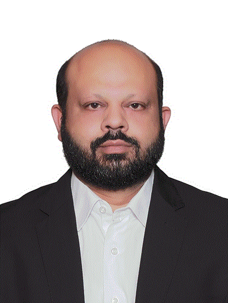
Syed Amir Gilani
The University of Lahore, Pakistan
Title: Sonography of the wrist examination technique
Biography:
Prof. Syed Amir Gilani is Founder Dean of the Faculty of Allied Health Sciences at University of Lahore, Lahore, Pakistan. He founded this faculty having 11 departments of Allied Health Sciences in 2012 which is having a student body of 5300 offering variety of courses including undergraduate to postgraduate degree programs. He is also a professor of radiology and director of Directorate of Global Linkages at the same institution. He received his MBBS from Punjab Medical College, University of Punjab, Pakistan, followed by DMRD & MPH from The University of Lahore and he holds doctoral degree (PhD) in Medical Diagnostic Ultrasound from Al-Zaiem Al-Azhari University (Sudan).Prof. Gilani is the author of 11 books on Ultrasound, has published more than 110 research articles in journals and has had above 320 international conference presentations & 81 conference publications. He has supervised 168 M.Sc, M.Phil 56 and 28 PhD Students from four universities of Asia, Africa & Europe. Furthermore, he has so far attended and conducted 128 conference/workshops in 80 countries.
Abstract:
Indications: Tendinitis-tenosynovitis, Tendon –rupture, Median Nerve compression-Carpaltunnel, Ulnar Nerve compression, Masses-ganglion-tumors, Synovitis, Joint effusion, US-guided aspiration, Loose Bodies, Bone erosions
Patient Positioning & Examination Technique - Hand and Wrist: Anterior scanning, Posterior scanning, The carpus or wrist. Note that its rounded proximal surface articulates with the radius and a fibrocartilaginous disk on the ulnar side. The carpus is concave anteriorly, forming the carpal tunnel which allows nerves and tendons to pass through to the palm. Ultrasound examination of the hand and wrist requires the patient to sit comfortably with their arm supported on an adjustable stand. The wrist is placed comfortably in a neutral position with a small rolled towel placed beneath it. The examiner performs the study facing the patient. Transducer: Ultrasound permits the performance of a dynamic examination as well as the ability to compare anatomy on the other side. Because the structures of interest are situated extremely superficially, a high frequency transducer in the 10- 12 MHz range should be used for examination.
Doppler: Doppler are also useful tools when available, especially for diagnosing hyperemic changes secondary to inflammation. Images are obtained in both longitudinal and transverse planes, following the natural course of the structures being examined.
Normal Ultrasound Appearances; The correct transducer position for imaging the flexor (upper image) and extensor (lower image) tendons of the wrist and sonography of various other normal structures will be discussed.
Pathologies: Carpal Tunnel Syndrome, Ganglion Cysts, Foreign Bodies (esp. radiolucent). Tendon tears (Flexors & Extensor),Tenosynovitis, Trigger Finger, Gamekeeper Thumb (UCL), Stener Lesion, Extensor Hood Injury, Pulley Tear, Pseudotumors Tumors. Foreign Body : Glass Rt 2nd finger PIP joint area, Tear: Thumb Extensor, Long Axis Rht EPB Tendon, De Quervain's Disease, Cronic Tenosynovitis-De Quervain,Trigger Finger (or Digit). Trigger digit is also a type of idiopathic tenosynovitis most often affecting the thumb, followed by the ring, middle, little and index fingers will be discussed. More commonly seen in women in the 40-60 year range, patients present with triggering or locking of the affected digit. Trigger Finger (or Digit), sonographically, a hypoechoic nodule is identified on the flexor tendon surface proximal to the A1 pulley. Fluid within the synovial sheath as well as synovial sheath thickening may also be present, often with small peritendinous cysts.
