Scientific Program

Conference Series Ltd invites all the participants across the globe to attend 2nd World Congress on Medical Imaging and Clinical Research Paris, France.

Day :

  • Radiography | Image Proceesing | Imaging Technology
Location: Sunset -1
Speaker

Chair

Abdulrahman A. S. Alsayyari

Qassim University, Saudi Arabia

Speaker
Biography:

Abdulrahman Alsayyari, is a vice dean of the college of applied medical sciences at Qassim university. He obtained his PhD degree from university of Queensland at Australia. He worked in both sector clinical and educational where he developed his experience and knowledge to improve the healthcare services for the Saudi population.    

Abstract:

The objective of the article was to study the common requested radiographs and relative exposure dose in Qassim province in Kingdom of Saudi Arabia. The method was retrospective and analytical study for collected variables as radiographs, relative entrance surface dose (ESD) and the effective dose, patient age, gender and causative factors. The doses have been derived from the product of system output, mAs, back scatter factor BSF, focal detector distance FDD and focus – skin- distance FSD based on the equation stated by ICRU, (2005) and  Davies et al, (1997):

The objective of the article was to study the common requested radiographs and relative exposure dose in Qassim province in Kingdom of Saudi Arabia. The method was retrospective and analytical study for collected variables as radiographs, relative entrance surface dose (ESD) and the effective dose, patient age, gender and causative factors. The doses have been derived from the product of system output, mAs, back scatter factor BSF, focal detector distance FDD and focus – skin- distance FSD based on the equation stated by ICRU, (2005) and  Davies et al, (1997):

Dose (mGy) = (Output(mGy⁄mAs)×(mAs)×(BSF)×〖(FDD)〗^2)/〖(FSD)〗^2

Then the effective dose in mSv has been derived from the equation stated by ICRP, 2007 report 103.

EffD=  ∑(W_T [H_T (female)+ H_T (male)])/2

Where WT refers to weighting factor for organ or tissue and HT refers to equivalent dose to organ or tissue.

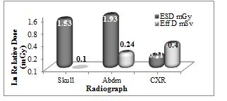
The analysis with excel software revealed that: the common requested radiographs were skull, abdomen and chest with male incidence as 75%, 72.2% and 64% respectively relative to whole sample. Traffic accident (71%) and fall-down (45%) were the most causative factors among male, female respectively, with injuries as skull fissure fracture (77%), and intracranial hemorrhage (23%). The skull radiographs noted among the age group of 11-21 years and peaking at 36% among the age group of 22-32 years. The requested abdominal radiographs appeared among the age group of 13-21 years; with frequency of (19%) and peaking at 30% among the age group of 22-30 years; with injuries as spleen ruptures (42%) and liver (27%). The chest radiographs observed among age group of 3-13 years; with frequency of 4% and peaking among age groups of 14-24 & 25-35 years old with frequencies of 19% and 21% respectively, and injuries as ribs fracture (55%), ribs dislocation (15%), pierced lung (20%) hemorrhage (10%). The average ESD for abdomen, skull and the chest radiographs were 1.93±0.8, 1.53±0.6 and 0.21±0.2 mGy, which were increase linearly following the aging, and the average effective doses were 0.24±0.1, 0.1±0.1 and 0.4±0.2 mSv respectively.

Image :
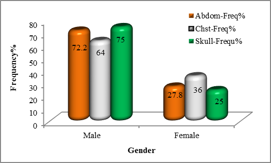
Figure 1: shows distribution of requested radiographic based on gender


Figure 2: shows the distribution of requested radiographic cases based on causes

Figure 3: shows the average ESD in mGy & EffD in mSv received by common anatomical site radiography

Recent Publications

1.      Kristin B Lysdah and BjÙ‘rn M Hofmann. (2009). What causes increasing and unnecessary use of radiological investigations? A survey of radiologists' perceptions. BMC Health Services Research, 9:155. doi: 10.1186/1472-6963-9-155.

2.      United Nations Scientific Committee on the Effects of Atomic Radiation. (2000). Sources and effects of ionizing radiation: report to the General Assembly, annex D, medical radiation

3.      ICRP. (1991). Recommendations of the ICRP publication 60, Annals of ICRP, Pergamon Press, Oxford.


Speaker
Biography:

Abdulrahman Alsayyari, is a vice dean of the college of applied medical sciences at Qassim university. He obtained his PhD degree from university of Queensland at Australia. He worked in both sector clinical and educational where he developed his experience and knowledge to improve the healthcare services for the Saudi population.    

Abstract:

The objective of the article was to study the common requested radiographs and relative exposure dose in Qassim province in Kingdom of Saudi Arabia. The method was retrospective and analytical study for collected variables as radiographs, relative entrance surface dose (ESD) and the effective dose, patient age, gender and causative factors. The doses have been derived from the product of system output, mAs, back scatter factor BSF, focal detector distance FDD and focus – skin- distance FSD based on the equation stated by ICRU, (2005) and  Davies et al, (1997):

Dose (mGy) = (Output(mGy⁄mAs)×(mAs)×(BSF)×〖(FDD)〗^2)/〖(FSD)〗^2

Then the effective dose in mSv has been derived from the equation stated by ICRP, 2007 report 103.

EffD=  ∑(W_T [H_T (female)+ H_T (male)])/2

Where WT refers to weighting factor for organ or tissue and HT refers to equivalent dose to organ or tissue.

The analysis with excel software revealed that: the common requested radiographs were skull, abdomen and chest with male incidence as 75%, 72.2% and 64% respectively relative to whole sample. Traffic accident (71%) and fall-down (45%) were the most causative factors among male, female respectively, with injuries as skull fissure fracture (77%), and intracranial hemorrhage (23%). The skull radiographs noted among the age group of 11-21 years and peaking at 36% among the age group of 22-32 years. The requested abdominal radiographs appeared among the age group of 13-21 years; with frequency of (19%) and peaking at 30% among the age group of 22-30 years; with injuries as spleen ruptures (42%) and liver (27%). The chest radiographs observed among age group of 3-13 years; with frequency of 4% and peaking among age groups of 14-24 & 25-35 years old with frequencies of 19% and 21% respectively, and injuries as ribs fracture (55%), ribs dislocation (15%), pierced lung (20%) hemorrhage (10%). The average ESD for abdomen, skull and the chest radiographs were 1.93±0.8, 1.53±0.6 and 0.21±0.2 mGy, which were increase linearly following the aging, and the average effective doses were 0.24±0.1, 0.1±0.1 and 0.4±0.2 mSv respectively.

Image :

https://d2cax41o7ahm5l.cloudfront.net/cs/upload-images/imaging2017-33417.png


Figure 1: shows distribution of requested radiographic based on gender


https://d2cax41o7ahm5l.cloudfront.net/cs/upload-images/imaging2017-17136.JPG


Figure 2: shows the distribution of requested radiographic cases based on causes


https://d2cax41o7ahm5l.cloudfront.net/cs/upload-images/imaging2017-17136.JPG


Figure 3: shows the average ESD in mGy & EffD in mSv received by common anatomical site radiography

Recent Publications

1.      Kristin B Lysdah and BjÙ‘rn M Hofmann. (2009). What causes increasing and unnecessary use of radiological investigations? A survey of radiologists' perceptions. BMC Health Services Research, 9:155. doi: 10.1186/1472-6963-9-155.

2.      United Nations Scientific Committee on the Effects of Atomic Radiation. (2000). Sources and effects of ionizing radiation: report to the General Assembly, annex D, medical radiation

3.      ICRP. (1991). Recommendations of the ICRP publication 60, Annals of ICRP, Pergamon Press, Oxford.


Speaker
Biography:

Masoud Hashemi MD, Anesthesiologist fellow in pain management has his expertise in pain management procedures in improving the health and wellbeing of chronic pain patients. He is known as the founder of the modern pain clinic in both educational and practical fields. His ten years of experience in teaching and training of physicians and so many patients who found less painful lives, prove his capacities in the field. As an associate professor and director of pain fellowship program and member of International Association for the Study of Pain (IASP) (Branch of Iran) he has a number of publications. Besides he organized and managed a number of congresses and workshops bringing out the latest and newest science based data as well as techniques in pain medicine in Iran.

Abstract:

There is a growing trend in ultrasound use in interventional pain management. Recently, the ease of use and clinical benefits of lumbar medial branch nerve block under ultrasound guidance have been identified. In this study, we assessed the relevant anatomy and sonoanatomy of these specific interventional techniques. We also evaluated the feasibility and success rates of ultrasound guided lumbar medial branch nerve block. Patients and Methods: Selected patients with facet joint pain who were referred to the Akhtar hospital pain clinics were evaluated.
Ultrasound-guided lumbar medial branch nerve blocks were performed. The target point for the lumbar nerve block was the cephalad margin of the transverse process groove in the vicinity of the superior articular process. C-arm fluoroscopy was performed to confirm the needle location. Pain levels were measured by a visual analog scale (0 - 10 scale), the Oswestry disability index (0 - 5 scale), and patient satisfaction scores (0 - 3 scale). The patients were followed for 6 weeks.
Results: The success rate was 98%, which was due to our use of ultrasound guided needle placement for the correct positioning of the needles. The mean procedural time was 5.9 ± 1 minutes. The average time of needle insertion was 4 ± 1 minutes. The pain intensity significantly improved from an initial value of 5 to 2.8 in the final follow-up (P = 0.0001). The oswestry disability index score significantly improved from 33.9 to 18.3 in the final follow-up (P = 0.0001). Patient satisfaction significantly improved from poor satisfaction immediately after the medial branch nerve block to excellent satisfaction in the final follow-up (P = 0. 0001). Analgesic requirements were also significantly reduced after 6 weeks of follow-up (P = 0.046).
Conclusion: Lumbar medial branch nerve block under ultrasound guidance was associated with high rates of treatment success and excellent treatment outcomes for facet joint pain. It is also feasible and administers no radiation. Thus, ultrasound-guided procedures can be used instead of conventional methods.

Biography:

 I completed my three-year diploma in medical radiography from Health Science School–College of North Atlantic–Qatar in 2008. Subsequently, I worked as a radiology technologist for Hamad Medical Corporation, Qatar, for two years. In 2010, I went to Edinburgh to continue my studies and obtain a bachelor’s in medical radiography from Queen Margaret University,Edinburgh. In 2014, I completed my bachelor’s and returned to Qatar, where I worked as a technologist. In 2016, I was promoted to technical supervisor at the National Center of Cancer Care and Research. I am always taking an active part in improving and developing my imaging skills, especially in pediatric imaging. In turn, I am sharing this knowledge as I train my colleagues and new staff in the department.

Abstract:

Autism Spectrum Disorder (ASD) characterizes as a mental disorder. According to Johnson et al., ASD is a developmental disorder of the brain that associates with impairments in social interaction, communication, and repetitive patterns of behavior; controlling their behaviors in usually challenging especially in hospitals. Johnson et al. show that children may become anxious in health care setup because of new faces of HCPs making them uncontrollable. Attending to such children would, therefore, need an experienced staff with good communication skills. Radiographers have a responsibility of ensuring smooth and effective communication with their patients to obtain a successful imaging. Mettler et al. present a research done in 2007 indicating that the amount of patients exposed to radiation has increased to a similar level to that of background radiation. It means that radiographers have failed in their responsibility hence putting the ASD children at more risk.
 
The objective of the research herein is to evaluate the efficiency of effective communication between radiographers and autism pediatric patient as a tool to reduction of radiation exposure.The study involves a qualitative research with two groups (n=10, five radiographers and five mothers to ASD children). The questionnaires were analysed by data analysis software, STRATA.
In results, mothers disclosed how their children behaved in different environments and what makes the children calm while radiologists expressed the challenges they face especially during imaging and gained strategies from mother's experiences. Good communication leads to easy and effective imaging procedure and thus, reduction in radiation dose in ASD patients.

Chih-Jen Hung

Taichung Veterans General Hospital, Taiwan

Title: MR myelography for postoperative orthostatic headache
Biography:

Chih-Jen Hung has his expertise in evaluation and passion in improving the health and wellbeing. He has worked both as Anesthesiologist and Pain Physician for a couple of decades. Based on the clinical experience of thoracic epidural analgesia for postoperative pain management, he tries to improve the practice of postoperative care and promote the education on the postoperative pain management.

Abstract:

We report the case of a man who presented with orthostatic headache postoperatively. The procedure of mid-thoracic epidural catheterization for posthoracotomy pain management was performed before induction of anesthesia. Unfortunately, theprocedure was abandoned due to some fluid aspirated from the epidural catheter. After operation, the patient complained of a severe headache for the first time when he was in the upright position. However, the findings of magnetic resonance (MR) myelography were characteristic of spontaneous intracranial hypotension (SIH), and the patient was successfully treated by an epidural blood patch at T1 level. Some reports implied that epidural anesthesia/analgesia might be a possible triggering factor of SIH in patients with an underlying structural weakness of spinal meninges. MR myelography can detect the pooling of CSF leakage. The T2-weighed MR image of our patient revealed dural sinus engorgement with contrast enhancement along bilateral nerve root sleeves of 5 levels from C7-T1 toT5-6 and left nerve root sleeve of C6-7 which indicated the presence of multiple simultaneous leaks at different spinal levels. The present case suggests that MR myelography could be carried out in patients with postoperative orthostatic headache when accidental dural puncture was not confirmed.

Image
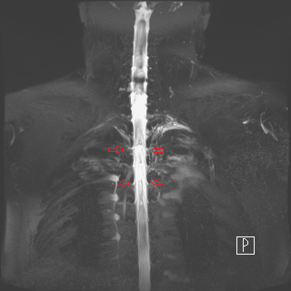
Figure 1: T2-weighed MRI revealed contrast enhancement along neural sleeves of C7-T1.

Figure 2: Many CSF leak lesions along multiple nerve root sleeves.

  • Medical Imaging | Radiology Trends and Technology | X-Ray and PET
Location: Sunset -1

Chair

M. A. Alnafea

King Saud University, Saudi Arabia

Biography:

M A Alnafea is presently working as an Assistant professor in King Saud University, Saudi Arabia. He attended several International and National conferences. He published several article in different journals as well.

Abstract:

This paper describes two non-Monte Carlo methods for investigating the possible application of Coded Aperture (CA) in breast tumor imaging. The first one based on a simple approach called Binary Mask Shift (BMS) representing the action of a distributed source in the projective CA imaging geometry. The second method based on Pseudo-Ray Tracing (PRT) that obtained by purely calculating the angle of incidence of each point in the object that successfully strikes an open aperture element and then hits the detector element. These methods particularly used for CA imaging investigations. Interestingly, these non-Monte Carlo methods yields similar results of a similar CA pattern but takes less computing power, than using a full MCS approach.

Biography:

Monica Kansal has been practising Radiology at eminent hospital and educational institutions from last nine years with special interest in Women’s imaging.

Abstract:

 Trans-vaginal sonography along with colour Doppler is the gold standard investigation in assessment of gynaecological and reproductive disorders in females. Besides exclusion of uterine, endometrial and tubal causes, sonography provides noninvasive tool for monitoring individual follicles during menstrual cycle and response to ovarian stimulation. This paper describes various uses of ultrasound in assisted reproductive techniques as the principal non-invasive modality for evaluation of key process of ovarian function – the process of folliculogenesis. Folliculogenesis refers to all phases that a primordial germ cell should pass to become mature healthy oocyte that is subsequently fertilized. It is a constant process that starts in embryogenic period and ends with the disappearance of last functional follicle in the period of menopause. Recognising the quality of follicle, its growth pattern and vascularity has a prognostic value for outcome of assisted reproduction techniques.
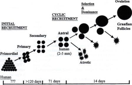
Figure 1: Folliculogenesis - Process of follicle recruitment and development.

Recent Publications

1.      V Vlaisavljevic and M Dosen (2007) Clinical applications of ultrasound in assessment of follicle development and growth. Donald School Journal of Ultrasound in Obstetrics and Gynaecology 1(2):50–63.

2.      Nanette Santoro, Barbara Isaac, Genevieve Neal – Perry, Tovaghol Adel, Laura Weingart and Aimee Nussbaum (2011) Impaired folliculogenesis and ovulation in older reproductive aged women. Journal of clinical endocrinology and metabolism: 88(11):5502–5509.

3.      Angela Baerwald, Gregg P Adams and Roger A Pierson (2012) Ovarian antral folliculogenesis during human menstrual cycle: A review. Human Reproduction Update, 18(1):73-91.

David Sipos

University of Pecs Faculty of Health Sciences, Hungary

Title: Radiographer prospects in Hungary
Biography:

David Sipos is a lecturer at the University of Pecs Faculty of Health Sciences Department of Diagnostical Imaging. According to my Bsc. degree I’m a radiographer,and I’ve done my Msc. from Healthcare Management. From September 2017 I’m going to attend the Phd. course at the University of Pecs. Next to my duties I’m also an application specialist for Medipixel Ltd., we are distributors of Samsung DR systems in Hungary and Slovakia as well. I can speak 4 languages (Hungarian,Slovak, Czech, English) and I’m highly interested in developing international relationships into the way of education and research.

Abstract:

Our aim was to identify the possible reasons behind the migration and attrition behaviour among Hungarian radiograhers. We were interested wether the dedicated professional staff has concrete ideas across borders which can be connected with attrition.For data collection we used one self made questionnaire and the Effort-Reward Imbalance (ERI) Questionnaire’s shortened version according to international practise. Our target group were workers with diploma of diagnostic analysis. Our sample counted 216
radiographers (n=216). For data analysis we used the 13.0 verison of SPSS. We applied analysis of variance, paired sample T-test, Chisquare test, linear regression and descriptive statistics with the performed probability of 95% (p=0.05). 30,6% of respondents (n=66) were concerned about the idea of working abroad, 40,3% (n=87) of respondents is staying in Hungary just because of their current life situation. At the group of 20-30 age we can espy significant relationship between the working opportunity abroad (p=0,001). The migration tendency was specific for radiograhers with no children (n=54). We found a significant relationship between the working ability abroad and the radiographers years spent in health care system (p=0,008). 41,7% of respondents (n=90) think that it will be favorable option if they change career. We found significant relationship between possible working opportunity abroad and the desire for higher wages and the lack of moving up in the radiographer hierarchy (p=0,001; p=0,001). Our research provides a broad view of the migration and attrition tendency among Hungarian radiographers. According to our study serious changes should be involved in the Hungarian system to keep the younger radiographers home.

Speaker
Biography:

Vikas Leelavati Balasaheb Jadhav has completed Post-graduation in Radiology in 1994. He has 23 years of experience in the field of Gastro-intestinal tract ultrasound and diagnostic as well as therapeutic interventional sonography. He has four Indian patents and an international patent in the field of Gastro-intestinal tract sonography and Radiology, since 2008. He has delivered many lectures in Indian as well as international conferences in nearly 27 countries as an Invited Guest Faculty, since 2000. He is a Consultant Radiologist and Specialist in Unconventional Gastro-Intestinal Tract Ultrasound and Diagnostic as well Therapeutic Interventional Sonologist in Pune, India.

Abstract:

Trans-abdominal sonography of the stomach and duodenum can reveal following diseases: gastritis, duodenitis, and acid gastritis. An ulcer, whether it is superficial, deep with risk of impending perforation, perforated, sealed perforation, chronic ulcer and post-healing fibrosis and stricture, polyps and diverticulum, benign intra-mural tumors, intra-mural haematoma,duodenal outlet obstruction due to annular pancreas, gastro-duodenal ascariasis, pancreatic or biliary stents, foreign body, necrotizing gastro-duodenitis, tuberculosis, lesions of ampulla of vater like prolapsed, benign and infiltrating mass lesions. Neoplastic lesion is usually a segment involvement, and shows irregularly thickened, hypoechoic and aperistaltic wall with loss of normal layering pattern. It is usually a solitary stricture and has eccentric irregular luminal narrowing. It shows loss of normal gut signature, enlargement of the involved segment seen, and shouldering effect at the ends of stricture is most common feature. Enlarged lymph-nodes around may be seen. Primary arising from wall itself and secondary are invasion from peri-ampullary malignancy or distant metastasis.
All these cases are compared and proved with gold standards like surgery and endoscopy. Some extra efforts taken during all routine or emergent ultrasonography examinations can be an effective noninvasive method to diagnose primarily hitherto unsuspected benign and malignant gastro-intestinal tract lesions, so should be the investigation of choice.

Biography:

Amjed Eljaili, MBBS, October 2010, University of Al-Zaiem Al-Azhari, Sudan, currently practicing in UK, Ysbyty Gwynedd, BCUHB, Intensive care unit, Surgical department Dr Eljaili has attended several academic meetings regionally and nationally also he has participated in various national work-shops, congress.
participation and membership with British institute of Radiology, United Kingdom.

Abstract:

The audit was carried out to exercise the compliance of the emergency department at Ysbyty Gwynedd, in relation to the standards guidelines of I-Refer and the royal college of radiologist (RCR), in order to ensure that the plain abdominal films are performed appropriately since it was noted that a significant numbers of abdominal X-rays are not clinically indicated or justified. Adherence to the recommended guidelines surely helps reducing the unnecessary radiation dose exposure directly toward sensitive and soft
abdominal organs and genitalia, also that will facilitate accessing more relevant imaging quicker into the way of establishing the diagnosis of the patient’s clinical problem. Abdominal X-rays is considered as high radiation dose study, and below is highlighting’s of few examples in comparison to the abdominal radiograph radiation dose,
1. Chest X-Ray: 0.1mSv (10 days background radiation).
2. CT head 2.0mSv (8 months background radiation).
3. Abdominal X ray 8.0mSv (3 years background radiation).
4. CT Abdomen and Pelvis 10mSv (3 years background of radiation).
5. CT chest 7mSv (2 years background of radiation).
The audit has also covered few other aspects in terms of film quality and request form adequacy as beside proper indication of the abdominal radiograph. The breakdown of the abnormal results illustrated by the radiology reports also has been mentioned, generally speaking, faecal loading represents the majority of that.
I-Refer/ RCR Standards guideline
The indications for requesting abdominal X-rays film, including:
1. Clinical suspicion of perforation or obstruction
2. Acute exacerbation of inflammatory bowel disease
3. Palpable mass (specific circumstances)
4. Constipation (specific circumstances)
5. Acute and chronic pancreatitis
6. Sharp/poisonous foreign body
7. Smooth and small foreign body, e.g., coin (specific circumstances)
8. Blunt or stab abdominal injury
Abdominal radiograph is not indicated routinely in the following conditions unless otherwise in very specific circumstances by specialist, pancreatitis, biliary Colic, constipation and GI Bleed.
Aim of the Audit: To audit our compliance in Ysbyty Gwynedd Hospital, emergency department, in adherence to the RCR/I-Refer standards guidelines and to increase the awareness of the healthcare staff in Ysbyty Gwynedd in relation to correct and appropriate indications of abdominal X-ray films, in so doing that will reduce to unnecessary radiation dose the patient will expose to. 2- To improve ‘the optimization of abdominal radiographic imaging in the emergency department in order to allow easy and correct interpretation of the radiograph film.
Methodology: This was a retrospective study involved data collection of 60 patients that were audited over period of two-monthsfor each cycle (Nov/December 2016 to March and April2017) to assess the compliance with the I-refer/RCR guidelines and also to optimise the film quality and the adequacy. The 60 consecutive abdominal X-rays requested in the emergency department where looked at their indication, radiology report, request form adequacy, radiograph quality and the clinical question need to be answered.
The patient’s information was obtained via using radiology information system for radiology reports/requests form and emergency department admission case cards.
Discussion and recommendations of the audit: It was noted that during the first the study, some abdominal radiographs were requested inappropriately for the following conditions, constipation, hemorrhoid, PR bleed, Upper GI bleed, ascites, bladder symptoms and UTIs, scrotal pain, appendicitis, gastroenteritis and acute cholecystitis, however majority of the films were requested by the most junior doctors particularly those who had recently completed a surgical placement. The junior doctors who newly started emergency medicine placement were educated and advised always not to hesitate to ask for senior opinion regarding requesting abdominal films when not sure whether or not indicated, also the majority of cases where bowels obstruction suspected have not had direct rectal examination performed as part for the NICE guideline for patients presenting with abdomenal pain.
Conclusion: Abdominal radiograph is of high radiation dose and usually has low diagnostic value in the majority of the cases, direct rectal examination is good practice and part of the recommended NICE guideline in the context of abdominal pain and bowel obstruction. Abdominal X-rays are not indicated for non-specific abdominal pain.

Biography:

Shahriari Mozhgan completed her study from Babol University, Iran. She has worked as a Researcher in the Radiology Research Center of Tehran University for six months and now she is working as a General Practitioner at her office.

Abstract:

Coronary computed tomography angiography (CCTA) is a beneficial method for detection of coronary artery disease. In this study,we investigated diagnostic accuracy and predictive value of vessel specific calcium scoring in detection of coronary stenosis by using 128-slice computed tomographic angiography (CTA) scanner. We used invasive angiography (IA) as the gold standard. 71 patients who had undergone both 128-slice CTA and IA were enrolled in the study. Three threshold for stenosis were considered (normal versus any kind of stenosis, stenosis<50% vs. >50%, stenosis<70% vs. >70%) in four major epicardial coronary arteries. Mean calcium score and p-values were compared between these three groups of stenosis by T-test and Mann-Whitney test. ROC analysis was done for evaluation of sensitivity/specificity, positive predictive value (PPV) and negative predictive value (NPV) of vessel specific calcium scoring method. There was a significant positive correlation between calcium score and coronary artery stenosis in our study.
The p value of this correlation for LAD in normal versus any kind of stenosis, stenosis<50% vs. >50%, stenosis<70% vs. >70% was 0.004, 0.005 and 0.001 respectively. For RCA, it was 0.001, 0.001 and 0.00 respectively and for LCX 0.02, 0.003 and 0.017 respectively.
In ROC analysis, we detected that by increasing in stenosis from normal to >70%, we had higher sensitivity, specificity and NPV in LAD, RCA and LCX arteries. Coronary artery calcification score is a good predictive and diagnostic method for coronary stenosis evaluation; however, it’s not enough in the case of high risk patients because it does not achieve 100% NPV.

  • Magnetic Resonance Imaging | Ultrasound | Clinical Research
Location: Sunset -1
Speaker

Chair

Daniel L. Farkas

University of Southern California, USA

Biography:

Hissa Nasser Mohammed completed his Diploma in Medical Radiography from Health Science School, College of North Atlantic, Qatar in 2008. He worked as
Radiology Technologist at Hamad Medical Corporation, Qatar for two years. In 2014, he completed his Bachelor degree in Medical Radiography from Queen
Margaret University, Edinburgh. He was Technical Supervisor at National Center of Cancer Care and Research.

Abstract:

Fluoroscopy is one of the radiation sources used in diagnostic processes in radiology. Owing to the diagnostic approach that entails observation of the affected anatomy using radiation in real time, harmful effects may potentially occur. Therefore, safety measures in the radiographer’s use of equipment and effective monitoring and management are essential in diagnostic processes. Additionally, patients in the age group of four to seven years have less anatomy and tissue development, which presents higher levels of health risks. The study will focus on analyzing the pre-procedure requirements and the set of guidelines, which enable the enforcement of the safety of the concerned patients within the pediatric practice. For example, the study will observe the potency of undertaking the reduction of the fluoroscopic times and improving the communications between the health specialists. The study will also evaluate methods and techniques employed in achieving efficiency in fluoroscopy radiation dose management. Direct and indirect methods will be employed to monitor the dosage effects. Direct methods would entail performing a skin dose test on the target area of fluoroscopic radiation. Detectors would be employed, for example the photographic films and thermoluminescent dosimeters. The indirect methods will employ the use of the dose area product meter to ascertain the effects of radiation on the patients. Some of the dose reduction techniques involve the manipulation of equipment operation, for example beam quality adjustment, dose level setting, and dose spreading. The results used to evaluate the dosage level will entail analyzing measurements of the skin exposure unit of the fluoroscopic equipment. Additionally, results from the change in beam quality and the effect on the skin will be analyzed in the study. These results will be achieved through the use of different operational voltage levels on the fluoroscopic equipment.
Skin dose will be determined through a combination of several measurable factors in fluoroscopic equipment operation. Due to the wide adoption of the fluoroscopic radiation technique in the pediatric oncology diagnostic process, dose monitoring is important in ensuring patient safety. Direct monitoring such as the skin dose procedure is effective, due to the reduced risk, which would otherwise result from other methods. Notable measures for efficiency in dose monitoring require adequate training of fluoroscopic operators on appropriate equipment use and continual observance of quality control procedures. These interventions and procedures should not compromise the quality of imaging and dose specification.

Speaker
Biography:

Daniel L Farkas is a former Fulbright scholar, directed a National Science & Technology Center at Carnegie Mellon University. He was Professor of Bioengineering at University of Pittsburgh, and Vice-chairman for research and Professor of Surgery at Cedars-Sinai Medical Center. His scientific interests center on investigating the living state with light, for uses in biology, bioengineering, medicine and surgery. He published 200+ articles and 28 books. He chaired 30+ international conferences.

Abstract:

Statement of the Problem: Alzheimer’s disease (AD) is a major unmet health challenge characterized by fast increasing incidence and costs.
Methodology: Our group has recently introduced optical imaging in the retina as a non-invasive method for mapping the occurrencesize and location of beta amyloid plaques, the primary pathology in AD. We have shown that using the fluorescence of curcumin,which attaches specifically to these plaques, we could quantitate the features of these plaques, including in vivo, and even document their reduction by immune treatments. These preclinical studies were also extended to the clinical domain, by using archival human eyes from patients with known levels of AD, as assessed both by brain histopathology and cognitive impairment (prior to death). We present a method for extending such studies to living patients, still using the retina as the window to the brain and plaques as indicators, but without the use of an extrinsic biomarker such as curcumin. This raises the level of experimental difficulty, thus requiring new technologies that we invented.
Findings: We designed a multimode optical imaging instrument, essentially a new type of confocal scanning laser ophthalmoscope,with some (needed) performance advantages over current commercial offerings. Our system consists of the following elements, all proprietary, and patent-protected: A highly versatile light source: pulsed, 400-1400 nm, with ~1 nm resolution; a new galvanometric method of scanning, with synthesized pivot point, not requiring a custom coupling lens; spectral analysis of imaging data, including
hyperspectral image segmentation and elimination of background; a more sensitive method of detecting light via parametric.
Conclusion & Significance: This new instrument achieves significant improvements in all of the following: spatial resolution,imaging depth, imaging angle in the retina (and thus spatial coverage), sensitivity and specificity. It will be used to image, fast and non-invasively, amyloid plaques in the retina, and any other retinal features of interest. We envisage that this instrument and the
approach it enables should be used in AD drug/treatment trials, as it allows the repeatable, non-invasive and quantitative imaging of amyloid plaques (via both their autofluorescence and scattering), and of their relationships with important structures in the eye, such as blood vessels.

Biography:

Claudia Paola Rivera Uribe has completed her medical school at the age of 24 years from Guadalajara University and posgrade studies from Nuevo León
Autonomous University. She is Chief of Residents of Pulmonary and Critical Care Medicine.

Abstract:

Invasive respiratory support is a cornerstone of Critical Care Medicine, however, protocols for withdrawal of mechanical ventilation are still far from perfect. Failure to extubation occurs in up to 20% of patients, despite a successful spontaneous breathing trial (SBT).We prospectively included ventilated patients admitted to medical and surgical intensive care unit in a university hospital in northern Mexico. At the end of a successful SBT, we measured Diaphragmatic Shortening Fraction (DSF) at the end of inspiration and at the end of expiration, and the presence of B-lines in five zones of the right and left lung. The primary objective was to determine whether analysis of DSF and Pulmonary Ultrasound improves prediction of extubation success. Eighty-two patients were included,24 (29.2%) failed to extubation. At univariate analysis, DSF (Youden’s J: > 30% [sensibility and specificity 62 and 50%, respectively])and number of B-lines zones (Youden’s J: > 1 zone [sensibility and specificity 66 and 92%, respectively]) were significant related to extubation failure (area under the curve 0.664 [0.526 to 0.801] and 0.819 [0.703 to 0.934], respectively). At the binomial logistic regression, only the number of B-lines zones remains significantly related to extubation failure (OR 5.91 [2.33-14.98], p < 0.001). In patients with a successfully SBT, the absence of B-lines significantly decreases the probability of extubation failure. DSF analysis does not add predictive power over the use of pulmonary ultrasound.

Speaker
Biography:

Ala Khasawneh is a Jordanian Doctor. He completed his Diploma in General Medicine (MD) and has been awarded the qualification of a Physician and title of
Doctor of Medicine from National Pirogov Memorial Medical University. He worked in Basma Hospital, Jordan. Currently, he is a PhD fellow in Diagnostic Medical Imaging in Hungary. As a Doctor, his main interest is to create new pathways for improving health care.

Abstract:

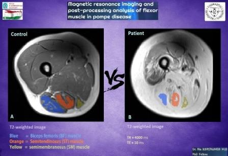
Pompe disease is a rare multisystem genetic disorder that characterized by a deficiency of the lysosomal enzyme acid alphaglucosidase and cytoplasmic glycogen accumulation causing damage that leads to muscle weakness. This study aim is to evaluate the muscle MRI pattern of twelve adults with late onset Pompe disease and twelve sex- and age-matched healthy controls (Age
range 19-59) for feature extraction which will be used to identify and classify functioning and non-functioning muscles. A training procedure was implemented using an Exercise Dynamometer device to stimulate the muscle for maximal contraction, MRI images data was used to compare the three flexor muscles in the lower limb function. MRI images data of biceps femoris (BF) muscle,Semitendinosus (ST) muscle, semimembranosus (SM) muscle before and after exercise (base, 30 min, 24 hours) was measured.
We performed and quantified T2 relaxation data of flexor muscles, and all data analyzed using repeated measure ANOVA to compare within related groups of the independent variable time (Base, 30M, 24H). According to our results, the significantly lower T2 value in the ST muscle of controls was observed (base=43ms, 30min=48ms, 24h=43ms; P < 0.05), but the change in SM muscle and BF muscle were not significant. In patients, we detected significantly higher T2 value in SM muscle evolve over time (base=129ms,30min=132ms, 24h=128ms; P < 0.05) compared to the controls, but ST muscle neither BF muscle doesn't show significant change.
As a conclusion, we can say that in Pompe patients the SM muscle can only react to the exercise apparently and shows us an activity in affected muscle cells, compared to the BF and ST muscle not shown any activity, that’s mean perhaps the Pompe disease change the muscle cells structure to interact to the exercise.
 

Syed Muhammad Anwar

University of Engineering and Technology, Pakistan

Title: Deep learning in medical image analysis
Biography:

Syed Muhammad Anwar is assistant professor at department of Software Engineering, University of Engineering, and Technology, Taxila and leading the Signal,image and multimedia, processing, and learning (SIMPLe) group. His research interest includes magnetic resonance imaging, machine learning, deep learning,medical image analysis and wearable and m-health.

Abstract:

Medical image analysis is the science of analysing or solving medical problems using different image analysis techniques for affective and efficient extraction of information. It has emerged as one of the top research area in the field of engineering and medicine. Recent years have witnessed rapid use of machine learning algorithms in medical image analysis.
These machine learning techniques are used to extract compact information for improved performance of medical image analysis system, when compared to the traditional methods that use extraction of handcrafted features. Deep learning is a breakthrough in machine learning techniques that has overwhelmed the field of pattern
recognition and computer vision research by providing state-of-the-art results. Deep learning provides different machine learning algorithms that model high level data abstractions and do not rely on handcrafted features.
Recently, deep learning methods utilizing deep convolutional neural networks have been applied to medical image analysis providing promising results. The application area covers the whole spectrum of medical image analysis including detection, segmentation,classification, and computer aided diagnosis. A brief introduction to the application of deep learning algorithms in medical image retrieval, segmentation, and detection will be presented.

Biography:

Kunwarpal Singh is Associate Professor in Department of Radiodiagnosis at Sri Guru Ram Das Institute Of Medical Sciences And Research, Punjab. He gave 13
publication and presentations in National and International Journals and conferences.

Abstract:

Background & Aim: Prostate cancer is one of the commonest malignancies in men that cause significant morbidity and mortality worldwide. The latest population-based cancer registry in India by ICMR records the age adjusted rate (AAR) to be 8.4, 10.7, 7.7,and 1.9 per 100,000 populations in the cities of Bangalore, Delhi, Mumbai and rural Barshi respectively. MRI is a powerful imaging modality for evaluation of anatomy and pathology of the prostate. This study aimed to evaluate the role of conventional MRI sequences
with diffusion weighted imaging using ADC values in staging of prostate carcinoma and in assessing response to treatment.
Materials & Methods: We prospectively studied 75 patients who came with suspicion of having carcinoma of prostate on Philips Gyroscan Achieva 1.5 Tesla MRI using sense body coil. Morphologic features of lesions were assessed on conventional sequences as well as on diffusion weighted imaging in pretreated and post treated patients. ADC values were calculated and statistically analysed.
Results: Highly significant correlation was found between serum PSA levels, prostate size, and staging and ADC values in both pretreated and post treated patients with p value <0.05. However, conventional weighted imaging did not show any significant change in signal intensity in post treated patients suggestive of added advantage of DWI.
Conclusions: MRI is a powerful imaging modality for comprehensive structural evaluation of anatomy and pathology of prostate
gland. Addition of ADC and DWI to T2WI provide more accurate results for prostate cancer detection, staging and post treatment
response.

Biography:

Shajeem Shahudeen has completed his MD in Radio Diagnosis from DY Patil University, Navi-Mumbai. He is the Managing Director and Consultant Radiologist at Vivid Diagnostic Centre, Kochi, India. He has also done several papers, poster presentation and publications.

Abstract:

Coronary artery disease (CAD) remains the leading cause of death in western countries with increasing prevalence in developing countries. The standard reference for diagnosis of CAD is coronary catheter angiography. Imaging of the heart has always been technically challenging because of the heart’s continuous motion.
CT imaging of the heart moved into the diagnostic realm by the introduction of multi-detector row CT (MDCT) and development of ECG-Synchronized scanning and reconstruction techniques.
These modalities allow for faster volume coverage, high spatial and temporal resolution. The introduction of MDCT especially has greatly benefitted cardiovascular CT applications as the speed of image acquisition shortens, breath hold and examination time for the patient and reduces the amount of contrast media needed for high and consistent vascular enhancement. The advents of 128-slice MDCT scanner sub millimetre resolution (0.4 mm) of substantial anatomic volumes are routinely achieved.
Aim of this study is: to study the role of MDCT coronary angiography in coronary artery disease (CAD) in symptomatic and asymptomatic patients; to study the calcium score in patients undergoing MDCT coronary angiography; to study the role of MDCT coronary angiography in patients with risk factors and to study the role of MDCT coronary angiography in follow ups of post-CABG and post-angioplasty stent patients.

Biography:

Dongyeon Lee completed his MS degree from Yonsei University, Republic of Korea, and is a PhD candidate in the Department of Radiation Convergence
Engineering at Yonsei University. He has published more than 10 papers in reputed journals and his research interests include CT, image registration, GPU
acceleration, and so on.

Abstract:

In conventional three-dimentional (3D) cone-beam computed tomography (CBCT) reconstruction, the image quality of reconstructed images is typically degraded due to patient’s motion such as respiration and heartbeat. In order to solve these difficulties, 4D CBCT incoporated with a phase-angle sorting scheme is often utilized. However, in this case, it requires dense projections in each phase to obtain reconstructed images of high quality and also severe aliasing artifacts are often present due to the limited number of projections at each phase used in the reconstruction. In this study, as an alternative, we propose an effective method for reducing motion blur and aliasing artifacts in conventional 4D CBCT reconstruction, the so-called deformable image registration(DIR), and made a quantitative comparison of the reconstruction qualities by both shcemes. In the proposed method, DIR process is carried out in two different ways as follows. In the first way, DIR process is applied to the resultant reconstructed images in 4DCBCT, while in the second way to the original projections on the basis of the reconstructed projections that are produced by forwardprojecting the reconstructed CBCT images in a single phase. The latter process is possibly performed using the adaptive steepestdescent POCS (ASD-POCS) algorithm for high reconstruction quality. Our results indicate that the motion blur and aliasing artifacts in the reconstructed CBCT images by using the DIR method were more significantly reduced, compared to the phase-angle sorting method. In addition, the second method in the DIR process seems more effective for reducing imaging dose than the first method.